


With Correlative X-Ray Micro-CT & X-Ray Fluorescence Microscopy
Abstract:
Worldwide, nearly 1 billion people are affected by nephrolithiasis, or kidney stone disease (KSD). Irrespective of age, sex, and race, a continuing increase in the international prevalence and incidence of KSD has been reported, with the annual direct and indirect cost of stone disease at an estimated $5 billion per year in the United States alone. KSD can lead to life-threatening infection and kidney injury, as well as chronic kidney disease and more systemic diseases, including diabetes, hypertension, and cardiovascular disease; furthermore, roughly half of all patients who experience a kidney stone will experience another episode within 5 years. While over 32 million Americans alone suffer from pane and quality-of-life inhibitions due to KSD, successful prevention requires a better understanding of the process of stone pathogenesis. In our research, we aim to increase our understanding of the mechanisms behind KSD development and biomineralisation, in order to develop definitive therapeutic approaches and preventative strategies [1-2].
Recent research has pointed toward the role of bacteria in stone formation, particularly in terms of understanding the formation of calcium-based calculi (accounting for ~80% of all KSD occurrences). It is our central hypothesis that urease-producing organisms such as P. aeruginosa, and CaOₓ -promoting E. coli, contribute to pathogenesis of stone formation by 1) nucleating new stones, and/or 2) interacting with existing stones. Our preliminary work has indicated that there are multiple bacterial species contributing to biofilm growth; thus, the focus of this research is on the commonalities between different stones in order to better understand the biomineralisation processes in urine-specific physiological conditions. Preliminary evidence has indicated that bacteria organise on and within the calcium crystalline material within the urinary system, thus the current research seeks to elucidate the mechanistic link between bacteria and the propagation of CaOₓ nephrolithiasis. [3]

Using a combination of high-resolution X-ray microscopy (microtomography / micro-CT) and X-ray fluorescence microscopy (micro-XRF), we have successfully visualised the complex 3-dimensional morphology of human-derived calcites. The XRF analysis, performed with the Sigray AttoMap-310, revealed the presence of both organic and inorganic materials, as well as a layered structure including the presence of Ca, P, O, and Mg. Furthermore, the presence of metallic minerals was also observed, most notably Fe and Zn.
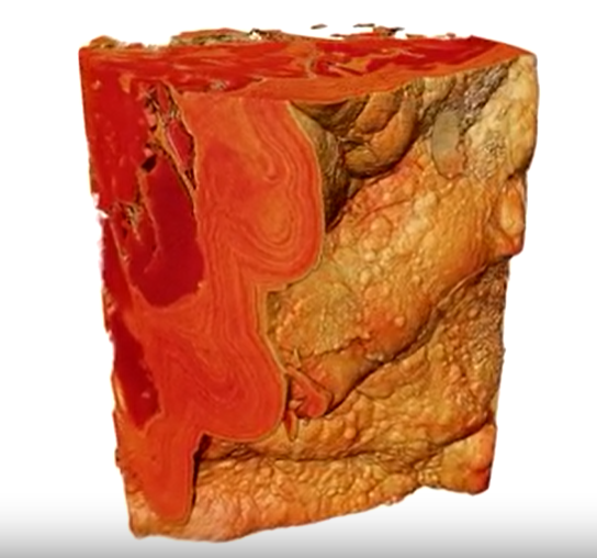
Pathologic human-derived calcite
Pathologic human-derived calcite imaged to show mineralogical differences at high resolution. Imaged using PrimsaXRM. Courtesy of UCLA Health
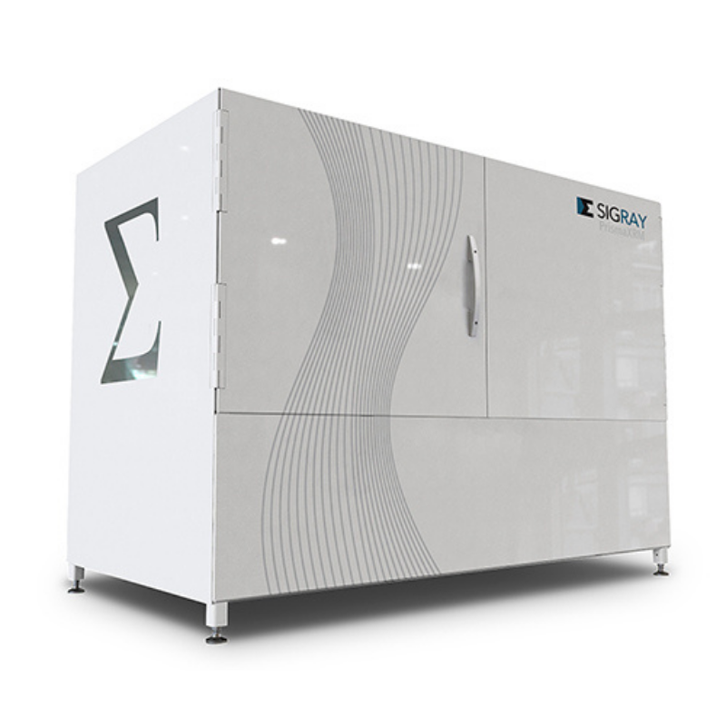
3D XRM analysis with the Sigray PrismaXRM-810 provided the spatial distribution of all of these materials, and revealed a complex 3-dimensional morphology including a layered architecture, with a uniquely-connected porosity present in each layer individually. With this combined x-ray microscopy technique, locations of bacteria may be identified as well as putative nucleation sites for stone formation. Through mineral classification via spectroscopic 3D signatures, we will demonstrate how two different types of X-ray analysis can be used together, providing a more comprehensive illustration of nephrolithiasis at the single-micron length scale than either technique used.

To discuss your application and the Sigray range of solutions, please contact our Technical Product Manager, Dr. Satyam Ladva by email or call (01372) 378822.
[1] Scales, CD et al. , Eur Urol 62 (2012), pp. 160.CrossRefGoogle Scholar
[2] Lotan, Y et al. , J Urol 172 (2004), pp. 2275.CrossRefGoogle Scholar
[3] An, L et al. , Oxid Med Cell Longev (2021).Google Scholar
Copyright © Microscopy Society of America 2022


