
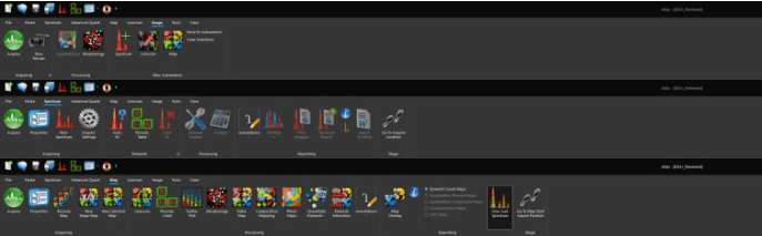
- Features
- Models
- Specifications
- Videos
- Downloads
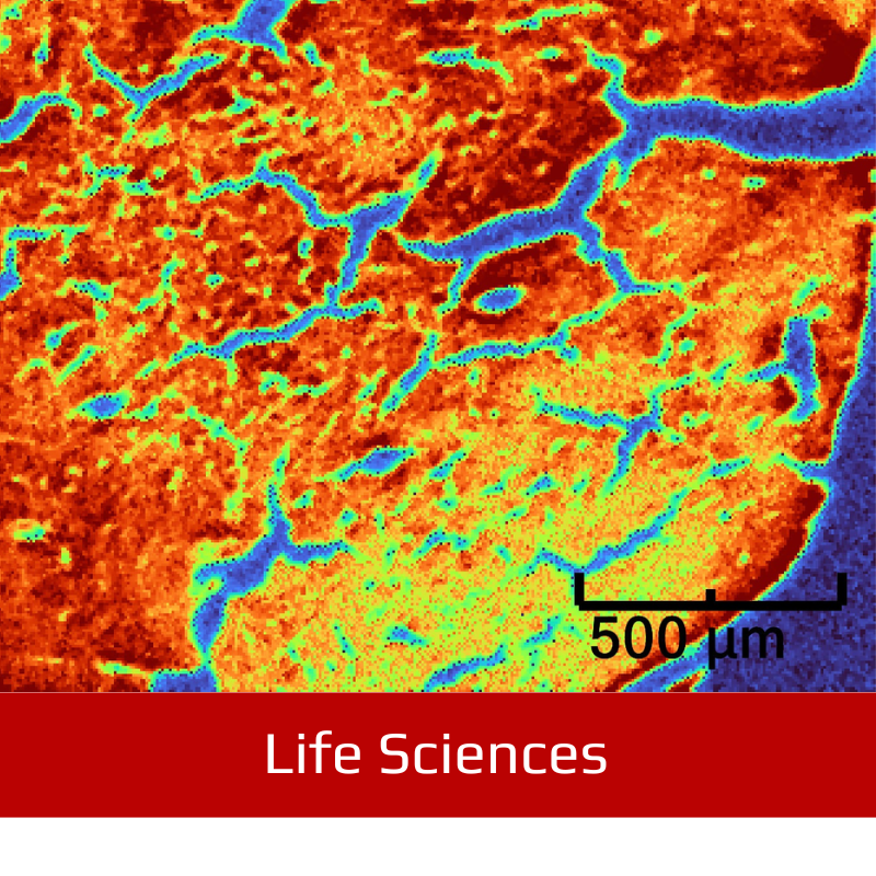
- Applications
- Related Products
- Contact
- Back To Spectroscopy
- Back To Optics
- Back To Hyperspectral
- Back To Cameras
- Back To X-Ray
- Back To Light Measurement
- Back To Characterisation
- Back To Electron Microscopy
- Back To Magnetometry
- Back To Ellipsometers
- Back To Cryogenics
- Back To Lake Shore
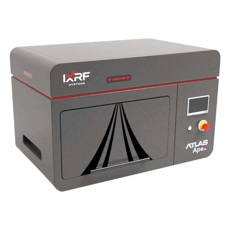
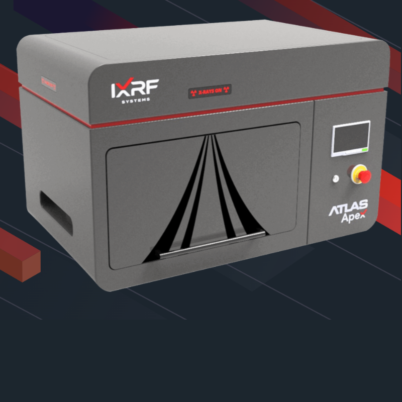
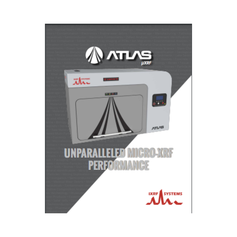
IXRF ATLAS Apex M : micro XRF Spectrometer
Micro X-ray Fluorescence (μXRF) Hyperspectral Imaging Elemental Analyser
Atlas Apex M is a benchtop microXRF spectrometer designed for non-destructive elemental mapping, hyperspectral XRF imaging, and quantitative micro-scale analysis of solids, liquids, particles, powders, thin films, and irregular samples. Built by IXRF Systems in Austin, Texas, it combines a high-flux microfocus X-ray source, polycapillary optics, perpendicular excitation geometry, configurable SDD detectors, and Iridium Ultra software to deliver advanced materials characterisation for research and industrial laboratories.
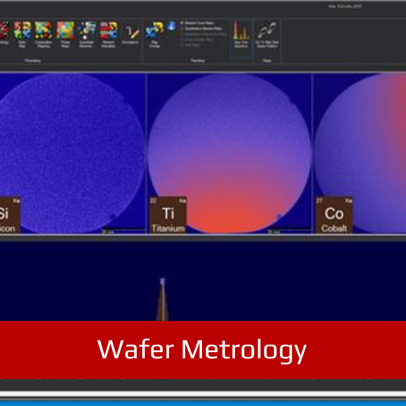
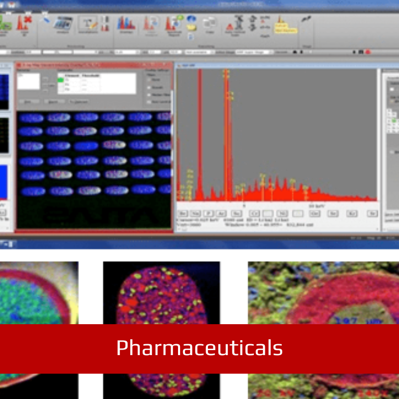
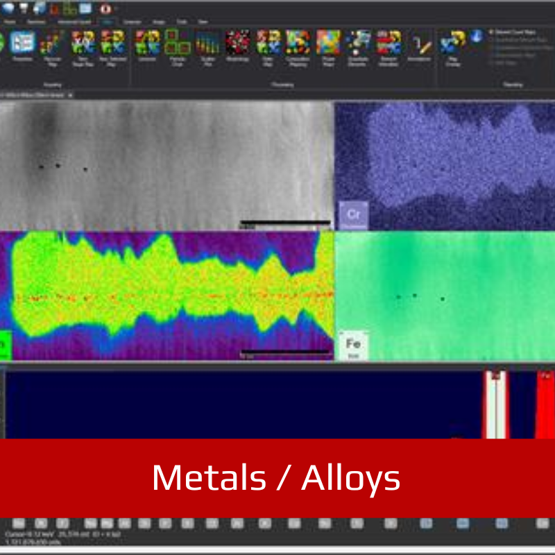
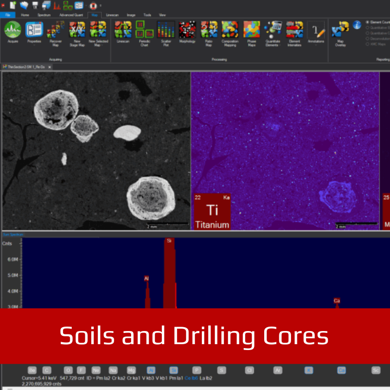
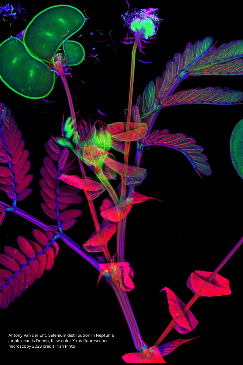
Engineered to visualise elemental distributions, identify small features, characterise phases, and quantify materials at the micro-scale, Atlas Apex M supports applications ranging from geology and critical minerals to semiconductors, batteries, coatings, metals, forensics, cultural heritage, environmental research, and advanced materials development.
ATLAS Apex M is the bench-top micro XRF tool of choice for sample characterisation using micro X-ray fluorescence (microXRF) hyperspectral imaging microscopy for information on composition (phase) and elemental distribution. With the smallest X-ray spot in the industry at 5 μm, this unique XRF microscope is optimised for analysis speed without compromising accuracy. The instrument can measure a wide range of sample types, whether small or large, even or irregularly shaped. Equipped with a large high-speed stage, it supports 2D analysis of virtually any kind of sample: solids, liquids, powders, thin-films and particles. Its large vacuum chamber delivers superior light element (low-Z) sensitivity.
FEATURES
- Benchtop / tabletop form factor
- Spot size down to 5 microns with anti-halo optic
- SDD detectors w/areas up to 65 mm2
- Large chamber volume
- 50 kV / 50 W polycapillary optic X-ray source
- Multi-point/multi-area automation & mapping
- Air, vacuum, helium for solids, liquids, and powders
- Iridium Ultra software running on Windows™ 11 OS
Profile

Profile
Profile

Profile

Profile

Profile

Profile

The 5 micron advantage
ATLAS Apex M is the bench-top micro XRF tool of choice for sample characterisation using micro X-ray fluorescence (microXRF) hyperspectral imaging microscopy for information on composition (phase) and elemental distribution. With the smallest X-ray spot in the industry at 5 μm, this unique XRF microscope is optimised for analysis speed without compromising accuracy. The instrument can measure a wide range of sample types, whether small or large, even or irregularly shaped. Equipped with a large high-speed stage, it supports 2D analysis of virtually any kind of sample: solids, liquids, powders, thin-films and particles. Its large vacuum chamber delivers superior light element (low-Z) sensitivity.
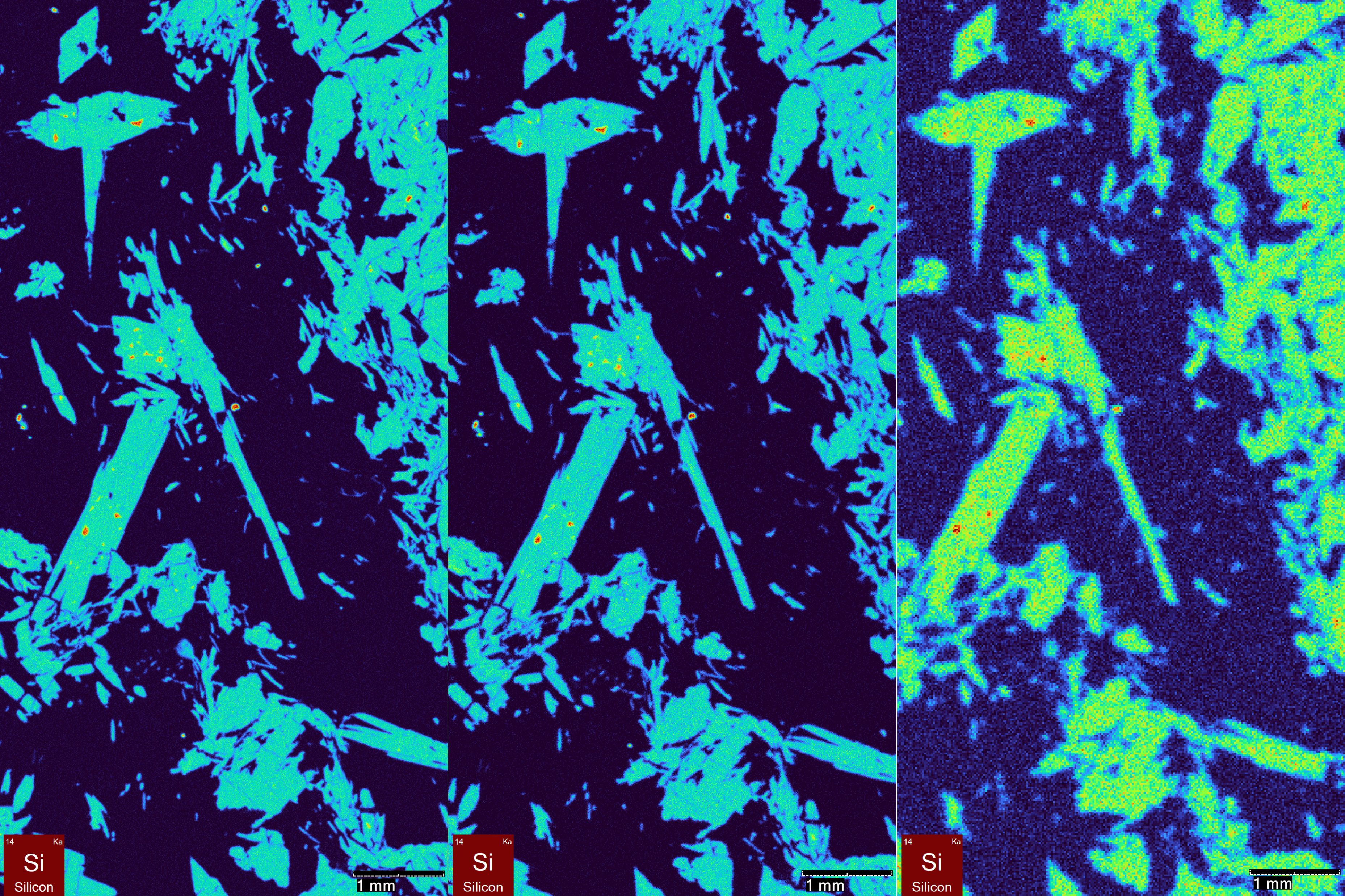
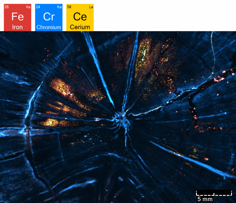
When it comes to XRF microscopic imaging (mapping), smaller is always better. Pictured here is a geologic thin section comparing (left to right) a 5µm beam with a 10µm and 20µm beam respectively.
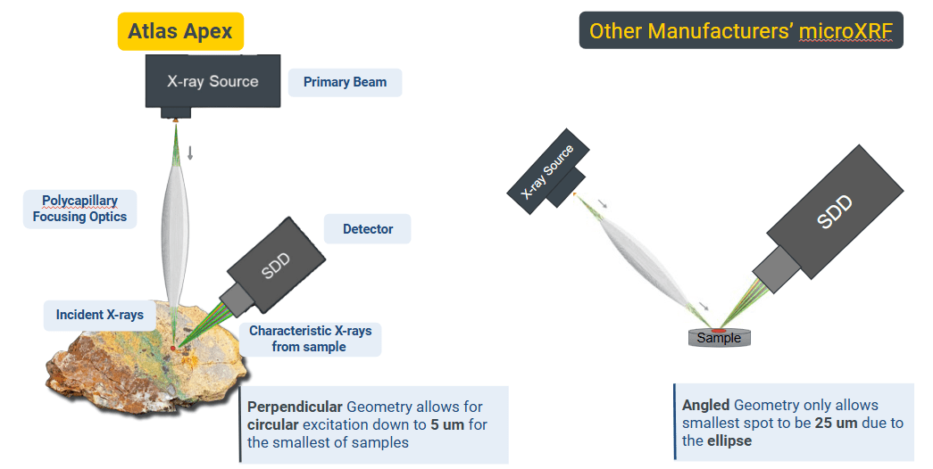
Superior geometry
- Perpendicular micro XRF geometry allows for circular excitation down to 5 μm for highest resolution.
- Beam is normal to the surface for most accurate hyperspectral imaging as beam is NOT an ellipse
- Competitor’s inferior angled geometry only allows smallest spot to be 25 microns due to the ellipse.
Unmatched speed
- Mix and match up to 4 Silicon Drift Detectors (SDD)
- For the largest possible solid angle collection efficiency
- Up to 260 mm2 area
- Energy resolution: ≤125 eV resolution
- Highest count rate with the smallest micro-XRF spot for fast high-res images
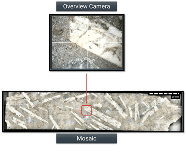
Mosaic automation
- Stitches high-magnification overview camera images into a single, large-area composite
- Enables full-sample visualisation beyond the camera’s native field of view (13×10mm)
- Supports acquisition at the highest available optical resolution for detailed inspection
- Facilitates efficient navigation and region selection for automated analyses
- Integrated with Atlas Automation to streamline sample setup and ROI definition
Spot automation
- Auto spectral spot analysis
- Automated microXRF mapping
- Automated linescans
Micro X-ray Excitation
- 50 kV / 50 W / 1 mA Rh target (other targets available) X-ray tube
- Polycapillary focusing optic
- Filter wheel with up to 7 filters (plus open position) before the focusing optic
- Available primary X-ray spot sizes: 5, 10, 15, 20, 25, 40, or 100 μm
- An optional second X-ray tube, with a choice of anodes, is available with spot sizes of 200, 500, or 1000 μm
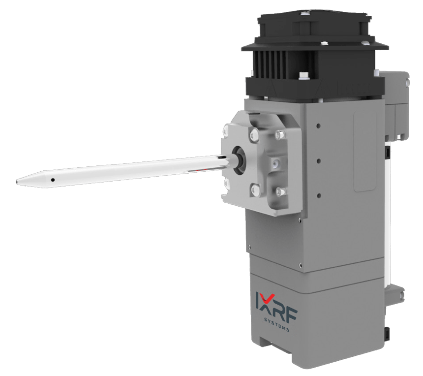
Detection
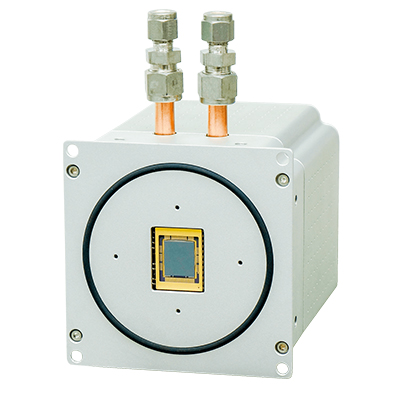
- Mix and match up to 4 Silicon Drift Detectors (SDD)
- For increased precision and/or reduced acquisition times
- 30 mm2 to 65 mm2 area
- Very close-coupled geometry
- Energy resolution: ≤125 eV resolution
- Peltier thermoelectric cooling
High precision stages
- Motorised X, Y, Z
- Speeds up to 300mm/s
- Map acquisitions at ≤ 1ms/pixel
- Accuracy < 1µm
- Custom adapters available
Iridium Ultra™ for Windows® 11
- Energy dispersive X-ray fluorescence (microEDXRF) spectroscopic software
- Fundamental Parameters (FP) for solids, liquids, powders, and particles
- Thin film FP for up to 8 layers, including an infinite base or substrate
- Automatic treatment for sum/escape peaks and full deconvolution
- Up to 8 acquisition conditions per analysis
- Hyperspectral EDXRF mapping and imaging, up to 9,998 × 9,998 pixels
- Every pixel is a full EDXRF spectrum
- Principal component analysis (PCA) for phase mapping
- IU-GEOCHEM (Geological Elemental Observation and Characterisation of High-Efficiency Mapping)
- Innovative mineral identification technology, comprehensive databases (4000 minerals) for quick and easy mineral identification
- Included with Iridium Ultra
Acquisition and quantitation
- One-click acquisition and automatic peak identification
- Customisable identification, labeling, processing, and quantification
- Scrolling periodic chart
- Drag and drop overlay
- Automatic overlap correction, sum/escape peak removal, background correction, and linear/non-linear deconvolution
- Fundamental Parameters (FP) and Quantitative Match
- Material classification database
- Empirical analysis via least squares and Lucas Tooth
Imaging and analysis
- Optical Mosaic Resolution: Ultra-High Definition Mosaic (Up to 100 Megapixels)
- Mosaic resolution is user-configurable and limited only by storage capacity and stitching parameters
- Multi-Point automated analysis directly from the image
- Morphological processing for rapid feature size measurements
- Image stitching and montage from the Overview
- Motorised zoom and focus control through software
- Live imaging during stage movements
- LED illumination with adjustable brightness
- Integrated crosshair overlay for accurate alignment of analysis points and mapping areas
- Imaging field of view matched to X-ray spot size for accurate correlation between optical and XRF data
- Segmentation and feature segregation
- Particle analysis package
- Including border removal, sorting, and exclusion
- Classification by composition
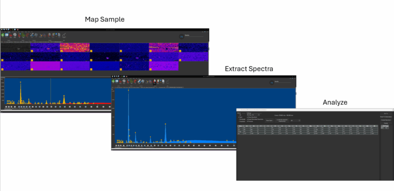
Mapping and linescans
- Simultaneous acquisition of 35 elements
- Full-spectrum hyperspectral imaging with up to ~100MP resolution
- Stored spectral data for every pixel
- Live spectrum display during acquisition
- Single or multiple map acquisition from image
- Overview or spot camera
- Map stitching and montage
- Extract spectra from map: point, area, freehand
- Create a linescan from map
- Mouse-over view intensities and concentrations
- Phase Analysis
- Multi-compositional map display
- Of element and compound ranges
- Overlay linescans on the image
Specialty and automation
- Multilayer thin film and coatings analysis up to 8 layers
- Serpentine continuous mapping (dual directional mapping)
- ASTM E2926-13 Glass Analysis
- Track, store, and recall all stage locations and images
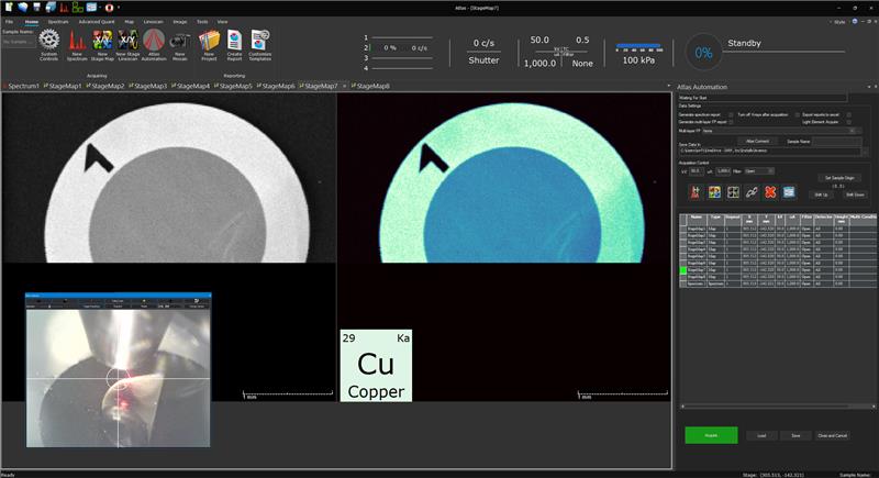
Imaging / mapping flow
- Create multiple X-ray maps on any region on the sample
- Define map parameters directly from video image
- Combine pixels to create analysis regions
- Regions are summed to create a composite spectrum
- Automatically identify elemental peaks
- Apply analytical quantification method
- Analytical results are displayed as a graphic
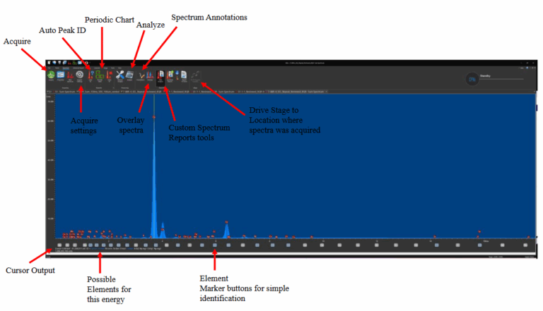
Top menu navigation
- Beginner users find the software simple to navigate
- Advanced users find that powerful tools are easily accessible
- All software features are included standard
- Custom report generation for completeness
- An exhaustive toolset comes from scanning electron microscopy
Spectral display features
- One click operation for acquisition, automatic peak identification, and quantification
- Collection of x-ray spectra into 25 individual windows
- Point and click cursor displays energy, counts, and possible elements present
- Customisable automatic element identification and labeling of peaks
- Scrolling markers from element chart on spectra
- Complete annotation tools for spectra; customise text, lines, and more
Mosaic navigation
- Stitches high-magnification overview camera images into a single, large-area composite
- Enables full-sample visualisation beyond the camera’s native field of view (13×10 mm)
- Supports acquisition at the highest available optical resolution for detailed inspection
- Facilitates efficient navigation and region selection for automated analyses
- Integrated with Atlas Automation to streamline sample setup and ROI definition
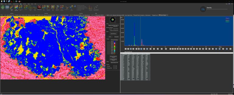
Automatic phase analysis
- Phase analysis by Principle Component Analysis (PCA)
- PCA is used in exploratory data analysis, an approach to analysing data sets to summarise their main characteristics
- PCA can be done on intensity or concentration based maps
- Phases are defined and percent (%) of field of view of each phase is defined
- In a PCA map, phases are marked by a defined color
- IU-GEOCHEM (Geological Elemental Observation and Characterisation of High-Efficiency Mapping)
- Innovative mineral identification technology, comprehensive databases (4000 minerals) for quick and easy mineral identification
Imaging composition
- User-defined compositions define the image
- Maps can be defined by intensities or concentrations
- Maps can be elements, compounds, components, materials, etc.
Line profile analysis (linescan)
- Relative elemental concentration or intensity along a line
- Multiple charts display the “tiled” form of the data
- Relative change of each element is viewed separately
- Linescans may also be displayed “stacked” for comparison
- Inset example shows an overlay of a long 300 mm linescan
- Larger image is a 5 micron scan at ppm levels
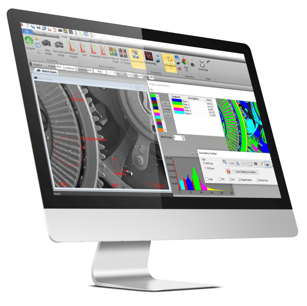
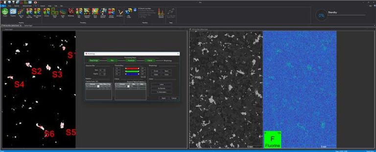
Morphology
- Surface morphology is a evaluation of the shape of a surface
- Particles, features, inclusions are elementally identified
- And automatically counted/measured by feature type
- Additional data on each feature may be easily collected
- As shown, an extensive set of image features is available
SPECIFICATIONS
- Elemental range: Fluorine (F) through Americium (Am). Carbon (C) through Americium (Am) on the LE version of Atlas
- Sample types: Solids, liquids, particles, powders and thin films
- Sample chamber size: 508 x 457 x 254 mm (20 x 18 x 10 inches)
- Analysis atmosphere: Air, vacuum or He(g) purge
- Primary X-ray source: 50 W max power, 50 kV @ 1 mA
- Optional secondary X-ray source: 4-12 W max power, 40-60 kV @ 0.4-1 mA
- X-ray source optics: Polycapillary or aperture collimation
- X-ray source anode: Rhodium (others optionally available)
- X-ray source spot size (primary): 5 μm standard (optional: 10, 15, 20, 25, 40 or 100 μm)
- X-ray source filters: Up to 7 plus an open position
- Primary X-ray source geometry: Top-down beam (perpendicular to sample stage)
- Detector(s): 1 standard, optionally up to a maximum of 4
- Detector types: Silicon drift detector (SDD) with Graphine windows
- Detector active area: 30 to 65 mm2, up to 260 mm2 w/ 4 detectors
- Sample stage type: Motorised X, Y, and Z
- Sample stage travel: 320(W) x 320(D) x 210(H) mm
- Mapping travel: 220(X) x 200(Y) mm
- Mapping scan speed: 1-3 ms/pixel
- Stage XY speed: Up to 300 mm/s
- Sample view: 3 cameras for sample positioning and analysis
- Operating system: SFF PC w/ Microsoft® Windows™ 11 OS
- Analysis and control software: Iridium Ultra: provides complete control of parameters, filters, cameras, optical microscopes, sample illumination and positioning, and measurement media
- Quality and safety: CE marked, RoHS, radiation < 0.5 μSv/h
- Dimensions: 890(L) x 560(W) x 560(H) mm (35 x 22 x 22 inches)
- Power: 100-240 V, 1 phase, 50/60 Hz