
- Features
- Models
- Specifications
- Applications
- Related Products
- Contact
- Back To Spectroscopy
- Back To Optics
- Back To Hyperspectral
- Back To Cameras
- Back To X-Ray
- Back To Light Measurement
- Back To Characterisation
- Back To Electron Microscopy
- Back To Magnetometry
- Back To Ellipsometers
- Back To Cryogenics
- Back To Lake Shore
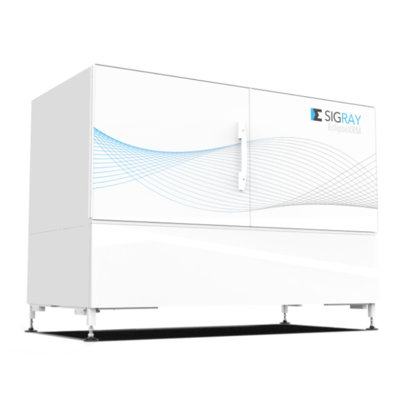
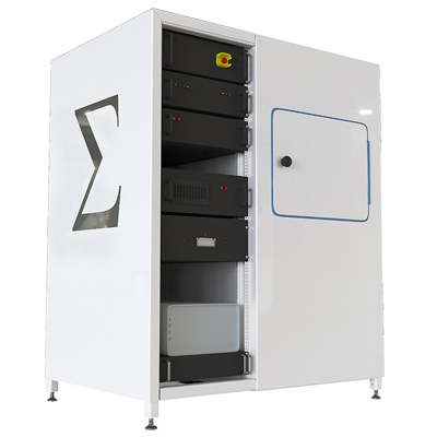
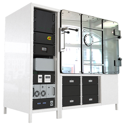
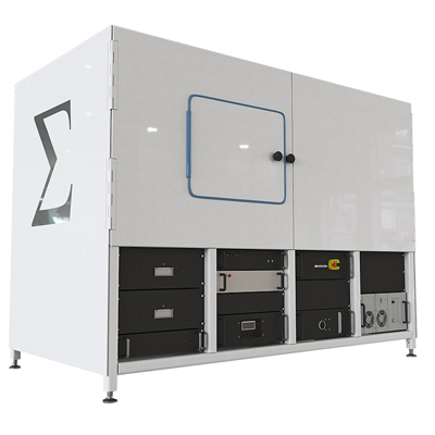
Sigray EclipseXRM-900 3D X-ray Microscope
Highest X-ray Resolution Performance
Patent-pending technology
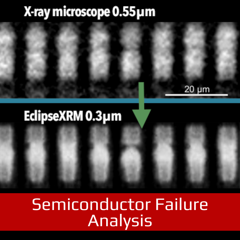
Spatial resolution of 0.3 µm
Submicron resolution, even for large working distances
EclipseXRM is a revolutionary system that bridges the x-ray microscope (XRM) gap between micro-XRM and nano-XRM, offering resolutions below 0.3 µm spatial resolution, even for large samples. This resolution far surpasses that of the leading flexible XRMs on the market, which are limited to ~0.5 µm spatial resolution.
The highest resolution achievable by the system is not only best-in-class, but is also achieved using a highly efficient detection system. This enables much higher throughput when imaging at smaller voxel sizes than other systems on the market.
FEATURES
- 3D x-ray microscope with breakthrough spatial resolution
Achieve down to 0.3 µm spatial resolution and <100nm voxels
- Maintain high resolution at large working distances
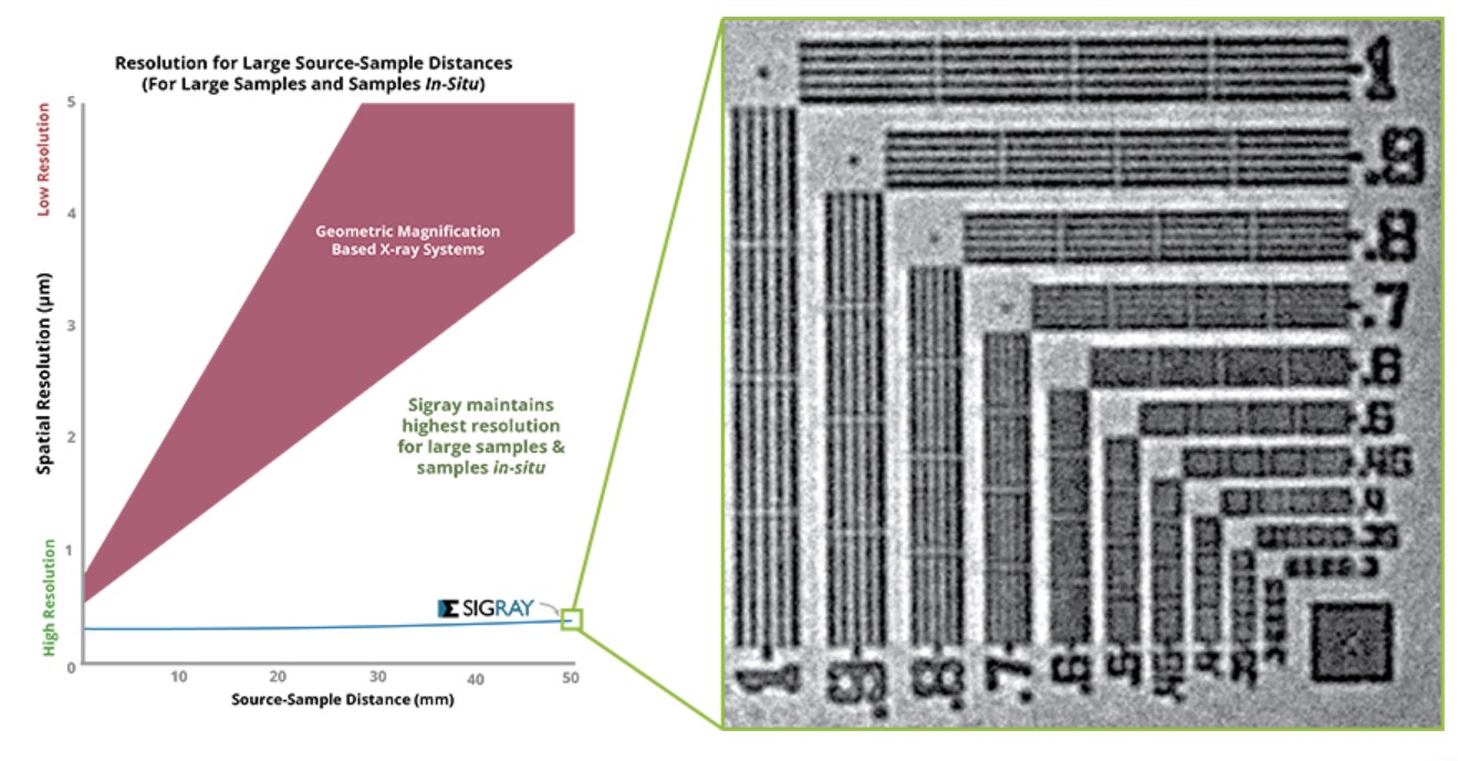
Submicron resolution achieved, even for large source-sample (working) distances. This is important for large or in situ samples
- Maximum versatility with novel contrast mechanisms
Patented Multi-Spectral Source (MSS) for quasi-monochromatic illumination to allow up to 10X throughput on the most difficult-to-image, low contrast samples
Profile

Profile
Profile

Profile

Profile

Profile

Profile

HIGHLIGHTS
Large samples and samples in situ typically require larger clearance around the sample for a complete rotation. This can therefore result in poor resolution on standard x-ray systems. One common strategy that has been heavily marketed to address this problem has been to use a “two-stage magnification” scheme using high magnification objective lenses. However, this has trade-offs in detector efficiency.
EclipseXRM’s new, patent-pending architecture offers an alternative to prior approaches, enabling submicron resolution at large working distances without needing high magnification objective lenses (see image).
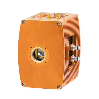
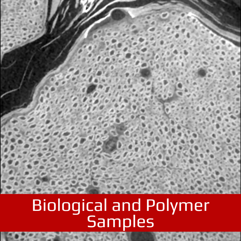
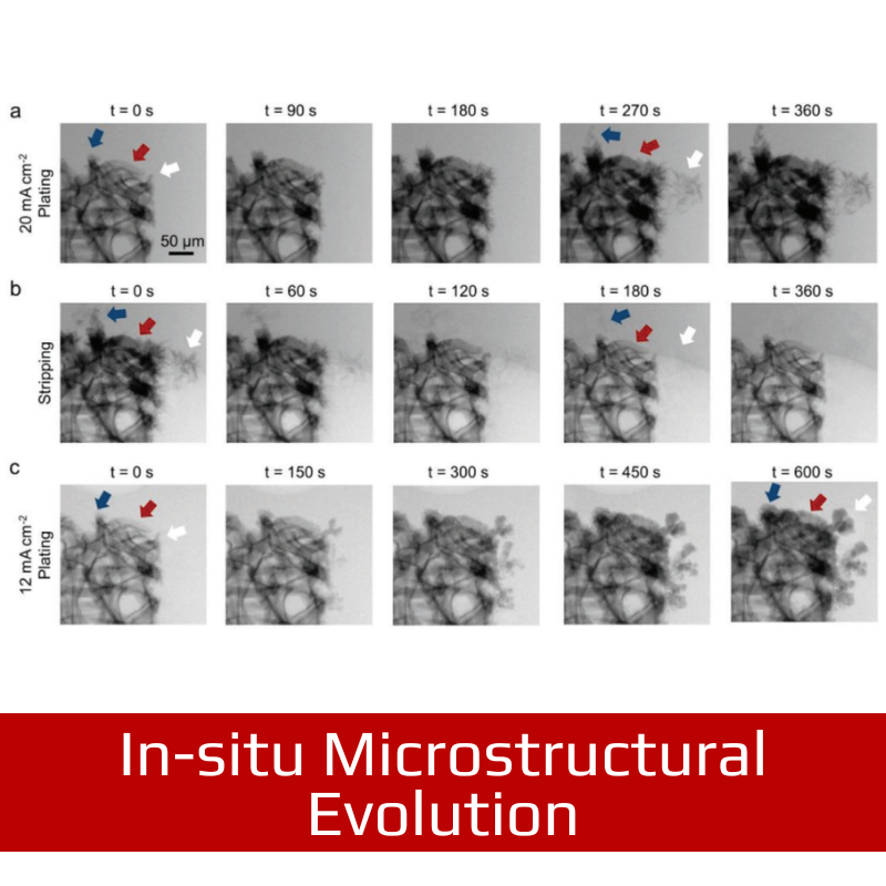
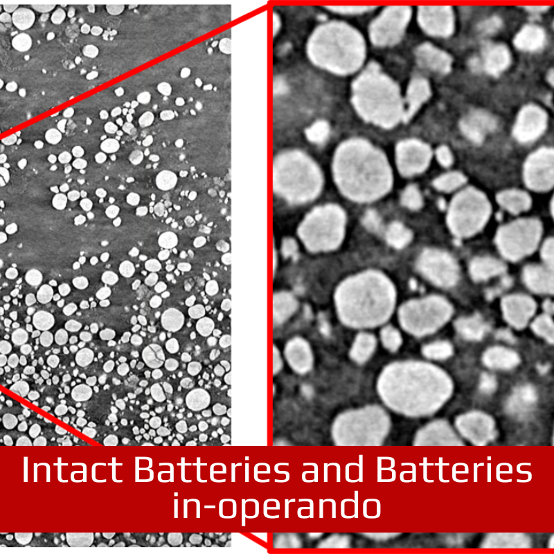
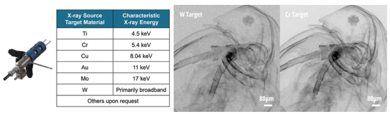
EclipseXRM was engineered to address the needs of busy central research facilities, encountering a wide variety of sample shapes, sizes, and compositions. These research facilities are often tasked with imaging the samples at the best quality and at the fastest acquisition times. As no single set of components can address the full range, EclipseXRM can incorporate multiple x-ray sources and detectors into a single system. One key option is a Multi-Spectral Source (MSS), which provides unique access to multiple quasi-monochromatic beams of x-rays (unlike the polychromatic x-rays produced by most system’s sources). This provides the outstanding contrast needed for extremely challenging samples, such as low-Z structures in novel lithium battery concepts and biological samples.
Image: EclipseXRM optionally incorporates a Multi-Spectral Source, which produces quasi-monochromatic x-rays for outstanding contrast for challenging samples.
EclipseXRM was designed by Dr. Wenbing Yun, who has won over four prestigious R&D 100 awards for his x-ray microscope innovations and has pioneered synchrotron and laboratory x-ray microscopes for the past four decades. The system takes advantage of several patent-pending and patented innovations that have been developed by Sigray, as well as the most recent hardware advances such as maintenance-free air-bearing rotary stages with sub-50nm runout and high-efficiency CMOS sensors.
The system has been adopted by some of the leading semiconductor companies and research institutions around the world.
SPECIFICATIONS
| Parameter | Specification | |
|---|---|---|
| Overall | Spatial Resolution | 0.3 µm |
| Resolution at Large Working Distances (100mm diameter sample) |
Submicron | |
| Contrast | Unique forms of contrast (phase contrast, Multi-Spectral Source) for biological, polymer, and geological imaging. Ask for the white paper. |
|
| Source | Source 1 | Nanofocus X-ray source 160 kVp Tungsten target on diamond |
| Source 2 (Optional) | Multi-Spectral Source (MSS) 100W, 50 kVp Up to 5 x-ray target materials. Includes selection from Cr, Cu, Rh, W, Mo, Au, Ti, Ag. Others available upon request. |
|
| Detector(s) | Types | Two detectors provided standard |
| Detector 1 | Large FOV (6.7 MP) HyperCapture-Pro CMOS Technology *Optional upgrades to 13 or 27 MP are possible. |
|
| Detector 2 | UltraVision-HDX Detector 10MP CMOS Technology |
|
| Software | Command and Control | Sigray 3D Features intuitive interface and AI for acquisition without requiring extensive training |
| Reconstruction | GigaRecon – fastest commercial CBCT reconstruction software (ask for our white paper) | |
| Offset Scans | Expands the horizontal FOV for ultrawide samples | |
| Helical Scan | Enabled for tall, cylindrical samples | |
| AutoPilot | AI-assisted microscope operation for unsupervised acquisition | |
| Linux Workstation | Interface is on a Windows workstation, while a separate robust Linux workstation controls the system. Advantageous for reliable 24-7 operation. | |
| EPICS | Open-source software controls for maximum flexibility and software extendibility | |
| Dimensions and System | Footprint | 100.8″ L x 53.6″ W x 85.3″ H |
| Stages | Maintenance-free air bearing rotary stage (recommended). Mechanical stages available upon request for rare use cases (e.g. facilities limitations). |