Researchers at Janelia Research Campus, Howard Hughes Medical Institute, are investigating the vast and complex network necessary for an organism to sense and react to the world. To do this, researchers must study neurons, which are the building blocks of the nervous system. Neurons connect to create complex networks at highly specialised junctions. Individual cells communicate at these ‘synapses’ by releasing chemical signals (or neurotransmitters) such as dopamine.
Despite the central role that synapses play in the brain, it remains challenging to measure exactly where neurotransmitters are released and how far they travel from their release site. Currently, most tools available to scientists only allow bulk measurements of neurotransmitter release. Using a Raptor Ninox II Vis-SWIR camera has enabled researchers to study how dopamine is released from subcellular compartments that have previously not been well characterised.
To tackle this limitation, the team at Janelia developed a new way to measure neurotransmitter release from neurons, harnessing a technique which uses fluorescent nanosensors that glow brighter when exposed to dopamine. These sensors form a very thin film upon which neurons can grow; when the cells release dopamine, the sensors ‘light up’ as they encounter the molecule. Dubbed DopaFilm, the technology reveals exactly where the neurotransmitter comes from and how it spreads between cells in real time. In particular, the approach showed that dopamine emerges from ‘hot spots’ at specific sites in cells; it also helped the team study how dopamine is released from subcellular compartments that have previously not been well characterised. Figure 1 highlights the schematic and workflow of the experiments.
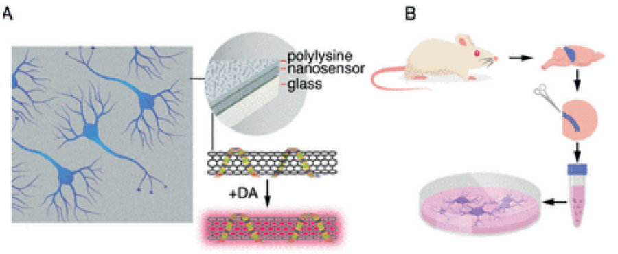
Figure 1: Schematic of DopaFilm imaging protocol. (see Ref [1])
(A) Schematic of DopaFilm.
(B) Workflow for preparing dopamine neuron primary cultures from the rat mid brain regions highlighted in blue. Neurons are grown on dishes with an engineered, chemisensitive, and fluorescent surface (DopaFilm) at the bottom.
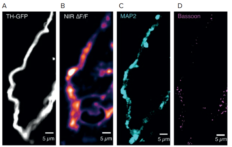
Figure 2: DopaFilm enables interrogation of the molecular correlates of dendritic release.
Activity from a dendritic process and its corresponding TH-GFP, MAP2, and Bassoon images. The NIR ΔF/F image contrast was set at 0–30% ΔF/F
DopaFilm requires use of detectors with InGaAs sensors for photon detection and additional optimisation of optical components to facilitate transmission of NIR and SWIR photons. Its nonphotobleaching fluorescence properties in the NIR to SWIR regions of the electromagnetic spectrum (0.85–1.35 μm) is spectrally compatible with existing optical technologies, as highlighted in figure 2.
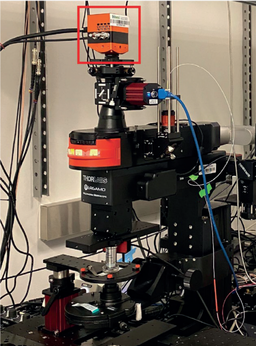
For broad-spectrum (visible to SWIR, 400–1400 nm) imaging, the team developed a custom microscope,
see figure 3, equipped with two fibre-coupled NIR lasers to excite far red, NIR and SWIR fluorophores:
a 671 nm and a 785 nm laser. The microscope is equipped with an Ninox 640-II camera with optimised
sensitivity in the SWIR range.
VIS-SWIR Imaging Camera.
Raptor has been developing InGaAs cameras for years. The Ninox family of cooled InGaAs cameras
enable longer integration times. These cameras are available in various resolutions and pixel sizes. As
well as being compact and low power, they provide high sensitivity, ultra-low noise images in visible and
SWIR wavelengths.
Ninox 640 II VIS-SWIR Imaging Camera.
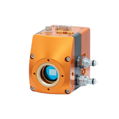
• VIS-SWIR InGaAs technology | Enables imaging from 0.6μm to 1.7μm
• Cooled to -15°C | Allows longer integration avoiding dark current build-up
• Ultra-Low Noise Sensor: 18e- in High Gain | Enables ultimate low light Vis-SWIR image
• 15μm x 15μm pixel pitch | Enables the highest resolution SWIR image
• Ultra high intrascene dynamic range | Enables simultaneous capture of bright & dark portions of a scene
• Onboard Automated Gain Control (AGC) | Enables clear video in all light conditions
• Ultra compact, Low power | Ideal for embedding into OEM systems
References:
Ref [1] “Visualising Synaptic Dopamine Efflux with a 2D Composite Nanofilm” https://elifesciences.org/articles/78773#fig5s7
Janelia Research Campus, Howard Hughes Medical Institute, United States; Center for Advanced Imaging, The University of Queensland, Australia

Any questions?
To discuss Raptor Photonics and your application, get in touch with our Technical Sales Manager, Dr. Luke Nicholls by email or call (01372) 378822.