


Micro XRF by iXRF has been utilised for several key areas in forensics crime scene analysis but also military grade techniques including
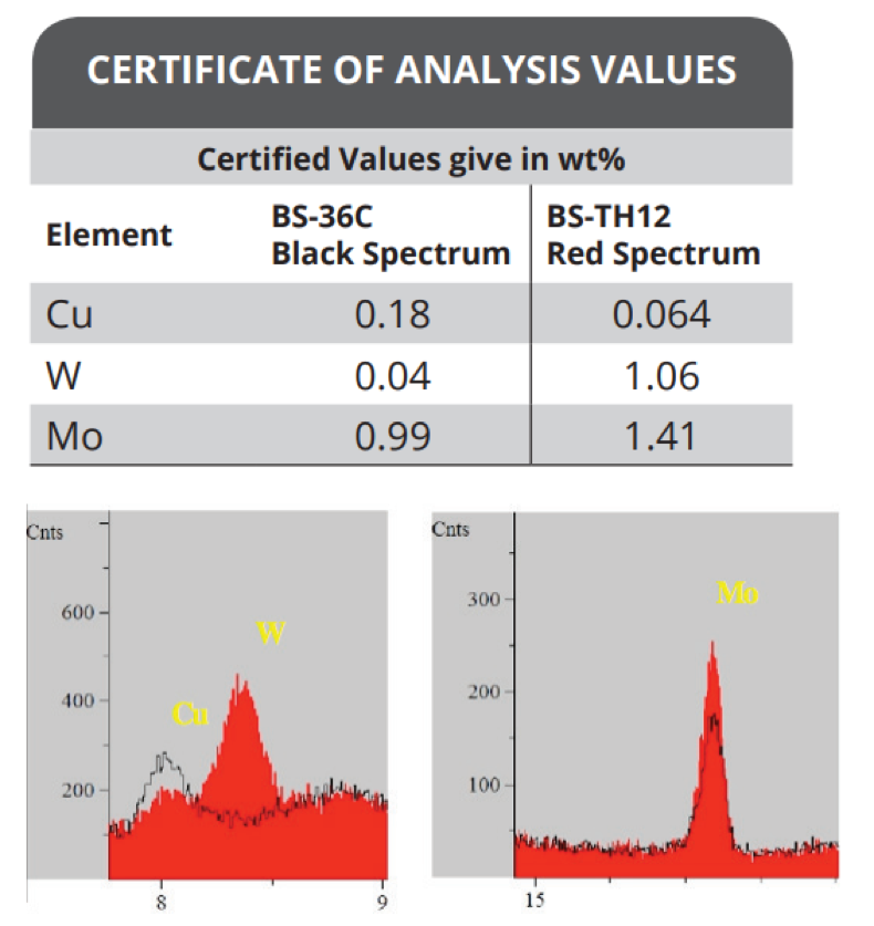
- Distinguishing between two very closely related steels to determine the nature of shrapnel from explosives such as pipe bombs through trace elemental analysis [Fig 1]
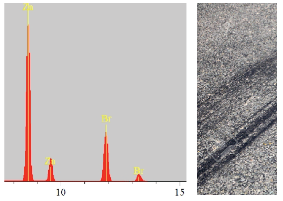
- Identifying scrapings from a tyre mark to determine make and origin of the vehicle [Fig 2]
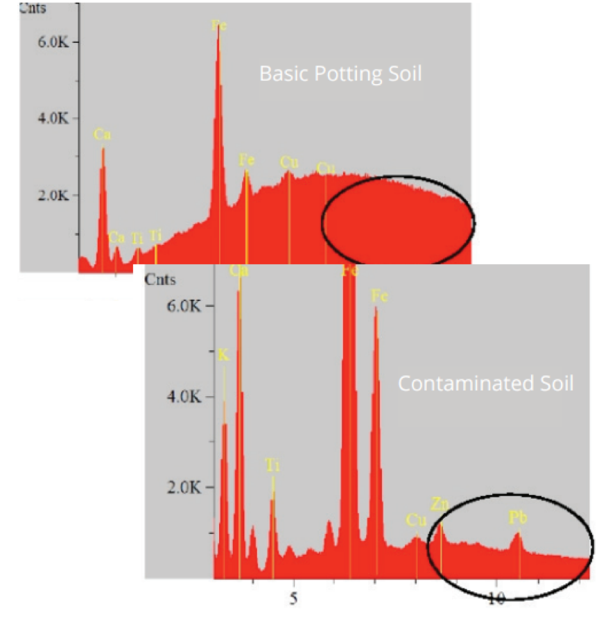
- Contaminated soil from underneath a shoe print can determine origin of suspect [Fig 3]
- Gun Shot Residue analysis can determine nature of projectile, muzzle to target distance and other parameters [Fig 4]
Scroll down to read more in depth application information on IXRF ATLAS and Forensics
Application Report
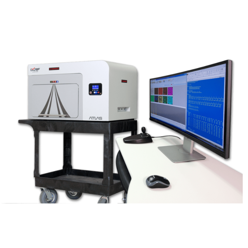
ATLAS and Forensics
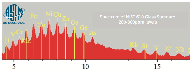
X-ray fluorescence (XRF) is a very useful and versatile tool to forensic scientists. It is a non-destructive technique, which is particularly advantageous if the sample is limited or further analysis is needed. Not only is it a non- destructive technique, it requires essentially no sample preparation. The ATLAS from IXRF Systems is a small spot μ-XRF unit which is capable of analysing extremely small samples down to 10 microns or scanning a small area of interest. μ-XRF is especially useful for analysing trace levels of elements in the ppm range, which can mean differentiation where other techniques falter or it could add to the weight of the evidence by showing samples cannot be differentiated.
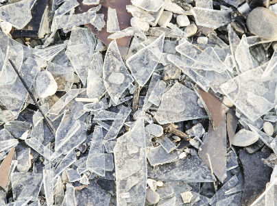
ATLAS can be used to analyse a wide variety of sample types, including but not limited to: glass chips, rubber, paint, soil/geological material, metals, money/coins, glitter, and more.
ATLAS is the only μ-XRF unit that has software that automatically processes and presents data according to the specifications in the ASTM method: “ASTM E2926- 13 Forensic Comparison of Glass using Micro-XRF Spectrometry”. The use of the software eliminates potential sources of human introduced error within the numerical data.
Paint Analysis
ATLAS is a very powerful tool capable of single or multi-spot analysis as well as elemental mapping to identify location of elements within a sample. This is extremely useful for paint chip/layer analysis.
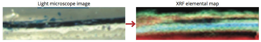
Due to the micron-sized spot of ATLAS different layers of paint chips are easily resolved not only elementally but also by comparative concentrations from one sample to the next. This can help differentiate closely related samples by the coating, paint, or base coat in any combination.
ATLAS can also be used as a bulk spot size XRF for homogenous evidence materials such as soil, metals, or other unidentified materials.
Comparative Analysis
Metal Comparison
XRF can help distinguish between two very closely related steel standards by comparing trace levels of elements. Shrapnel from a pipe bomb scene could be compared to metal shavings found in a suspect’s garage.
Soil Analysis
XRF can easily identify differences between common potting soil and contaminated soil. Soil found from a shoe print at a crime scene can potentially indicate the suspect has been to or is from a specific region
Tire Rubber Analysis
Identify trace elemental similarities or differences between tire mark and tire. Scrapings from a tire mark at a hit and run scene can be compared to a suspect vehicle.

Application Note: GSR on Textile
… using ATLAS microEDXRF Spectrometer
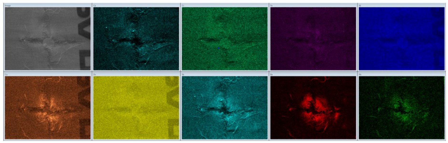
When a firearm is shot, in addition to the projectile(s), a mass of debris comes out the muzzle. These gunshot residues (GSR) can include various primer residues, residues from projectiles, and partially burned and unburned gun powered particles. The examination and analysis of GSR on items of evidence can allow determinations to be made as to whether a hole or defect is consistent with being caused by a bullet (or other rearm-related projectiles). In a crime lab equipped with an IXRF ATLAS series microEDXRF (micro-XRF) spectrometer, a forensic scientist can examine patterns of GSR on items of evidence to determine muzzle-to-target distance and other parameters of interest.
Gun shot residue fabric piece was mounted on a sample plate. It was elementally mapped in an area around a hole, with the X-ray tube set to 50 kV and 1000 μA (no tube filter). Map size was set to be 88.18 x 65.25 mm. Due to the sample not being perfectly flat when mounted, it was mapped with the stage slightly lower than the proper working distance to account for the variation in sample height. IXRF System’s ATLAS micro-XRF imaging spectrometer has the X-ray source pointing straight down onto the sample, which allows the user to alter the spot size to be larger as the stage is lowered from the optimal working distance.
This sample was imaged with a spot size of about 60 μm and was collected at optimised conditions to highlight the features of the unit, the spot size, and the software. The optimised map was collected for 35 hours. It is possible to collect a much faster map of the area of interest by simply altering some of the collection parameters. Optimised maps are shown below, with each square representing a different element. The elements are listed in the upper left corner of each map. Maps are from left to right, top to bottom: X-ray image, Ca, Cr, Fe, Ni, Cu, Zn, Sb, Ba, Pb.

*The text seen in the x-ray image as well as a few elemental maps is from the sample stage.
Image, Sb, Ba, and Pb maps from the previous screen are shown enlarged to show the detail collected in the maps.
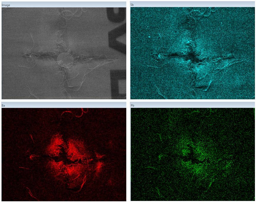
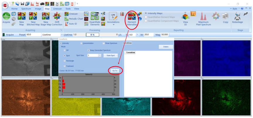
Elemental maps are shown as intensity, hence brighter areas are higher intensity. To compare the variation of the elements across the area of interest, so ware features were used. With the tool ‘Element Intensifies’ a spot can be selected on a map to either extract data from the map or to drive the stage to that exact spot on the sample, for further investigation, by using the ‘Go To’ button.
The ‘Go To’ feature was used to move to several locations to collect spectra for further comparison.
The selected spots are shown in the x-ray image and elemental map below. There is also a micron bar to reference size.
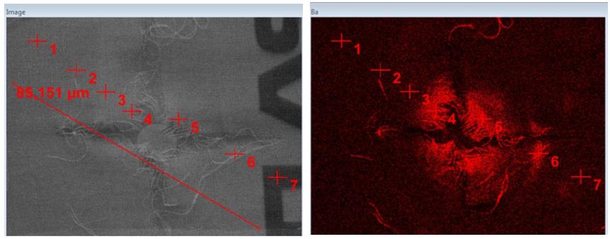
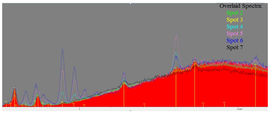
The resulting spectra collected from the seven spots are overlaid below. In the overlay Spot 1 is the red spectrum. The other spots are shown as the colours listed below.
The elements elicited were K, Sb, Ca, Ba, Fe, Cu, Zn and Pb.
The Rh is from the anode of the XRF and is expected to be in the spectrum. The Ar is from the air, as the spectra were not collected under vacuum.
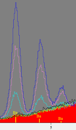
The Ba L-line region was magnified to highlight the variation in intensity for the different spots.
A portion of the fabric, not near the hole, was analysed to be used as background for comparison of which elements are from the GSR and which are from the fabric.
In the overlay below the red spectrum is of Spot 6 and the black overlay is from the fabric.
The fabric appears to contain Ca and a small amount of Fe. While the region considered to be GSR contains K, Sb, Ba, Fe, Cu, Zn, and Pb.
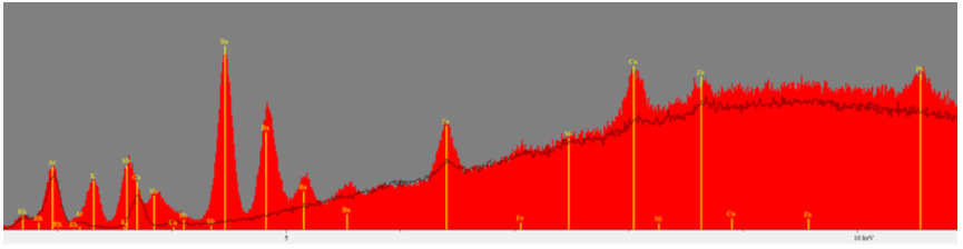
IXRF System’s so ware can use the spectrum from the fabric as background removal. The spectrum below is Spot 6, with the fabric spectrum/background removed.

Related Products
Sigray PrismaXRM Tri-Contrast X-Ray Microscope
Award-Winning Sub-Micron 3D X-Ray Microscope

Specim IQ Hyperspectral Imaging Camera
Portable carry on Hyperspectral Imaging Cameras
Contact

What’s your forensic application? To talk about IXRF system capabilities, get in touch with Dr. Satyam Ladva on (01372) 378822 or email.


