


Extract real 3D topography on hard to reach sample areas with subnanometer resolution and correlative SEM imaging.
Introduction
Bone tissue is continuously remodeled through the concerted actions of bone cells. Whereas osteoblasts synthesise the bone matrix and are responsible for its mineralisation, osteoclasts break down bone matrix. The third cells, osteocytes, are inactive osteoblasts that have become trapped within the bone they have formed. The small space containing the osteocyte is called lacuna. When the tissue is dead, the lacuna topography gives information about the osteocyte that occupied it before.
Scanning electron microscopy (SEM) can help to identify lacunae in bone structure. However, quantitative height information is not available in SEM. Atomic force microscopy (AFM) is an established tool for quantitative analysis, however, it is very difficult to access lacunae without optical guidance. Therefore, it is typically a very challenging task to identify the lacunae structures and position the AFM cantilever directly on the area of interest.
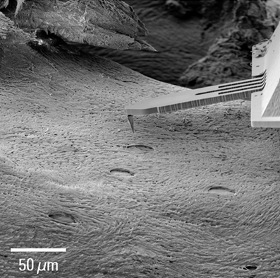
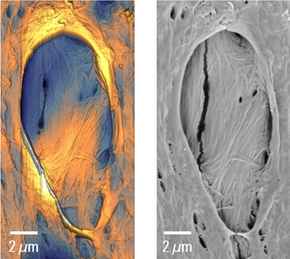
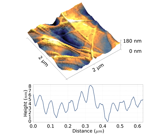
Figure2: FusionScope and Profile View allow an easy and fast positioning of the cantilever tip on a lacunae structure (Top). The joint coordinate system enables the extraction of correlative AFM topography (Bottom, Left) and SEM information (Bottom, Right) of the lacunae and corresponding collagen fibres.
Data Analysis
The FusionScope is a dedicated solution where both imaging modalities are seamlessly integrated within one instrument. Therefore, the user can combine the complementary strengths of both AFM and SEM. This is particularly advantageous when the sample area is extremely difficult to reach with the AFM cantilever as in the case of bovine vertebra. SEM’s large field of view enables the easy identification of lacunae in bone tissue and the high precision of the SEM, when used in conjunction with FusionScope’s Profile View, can then be used to position the cantilever directly onto a lacunae. With the help of the AFM the user can then extract quantitative 3D topography of the lacunae structure and is able to directly correlate it with the SEM image.
The FusionScope microscope allows for complementary SEM and AFM topography information of lacunae and collagen fibers. Real 3D representation of the topography can be used for analysing collagen fibers with a much higher resolution, allowing quantitative analysis of the characteristic periodic bending pattern on collagen fibers with sub-nm resolution. The cross section of the bending pattern shows a corrugation height of 2-3nmthat can be easily resolved with the FusionScope system. In summary, the FusionScope is the solution of choice to easily perform correlative AFM & SEM analysis. It accepts a large variety of samples and provides a wide range of different measuring modes. SEM guidance makes the identification and cantilever positioning very easy and lets you combine quantitative AFM data with all of your SEM analysis.
Easy to use Correlative AFM/SEM Microscopy Platform
Combine the complementary strengths of AFM and SEM like never before! The FusionScope fully integrates a wide range of advanced AFM measurement techniques with the benefits of SEM imaging. Seamlessly image your sample, identify areas of interest, measure your sample, and combine your imaging data in real time.

Interested to learn more about the FusionScope’s capabilities and how it could help with your next project?
Get in touch with Luke Nicholls by email, or call (01372) 378822.


