


Understanding magnetic phenomena at the nanoscale is crucial to advancing a wide range of scientific disciplines—from condensed matter physics and spintronics to emerging quantum materials. High-resolution magnetic imaging plays a vital role in revealing how magnetic domains behave and evolve within complex material systems.
In the newly released application note, “Magnetic Imaging with FusionScope®” researchers are introduced to the capabilities of the FusionScope®, an integrated platform that combines Scanning Electron Microscopy (SEM), Atomic Force Microscopy (AFM), and Magnetic Force Microscopy (MFM) in a single high-vacuum system. This note provides an overview of how FusionScope facilitates precise, correlative imaging of magnetic micro- and nanostructures with enhanced sensitivity and spatial resolution.
Magnetic Force Microscopy: A Brief Overview
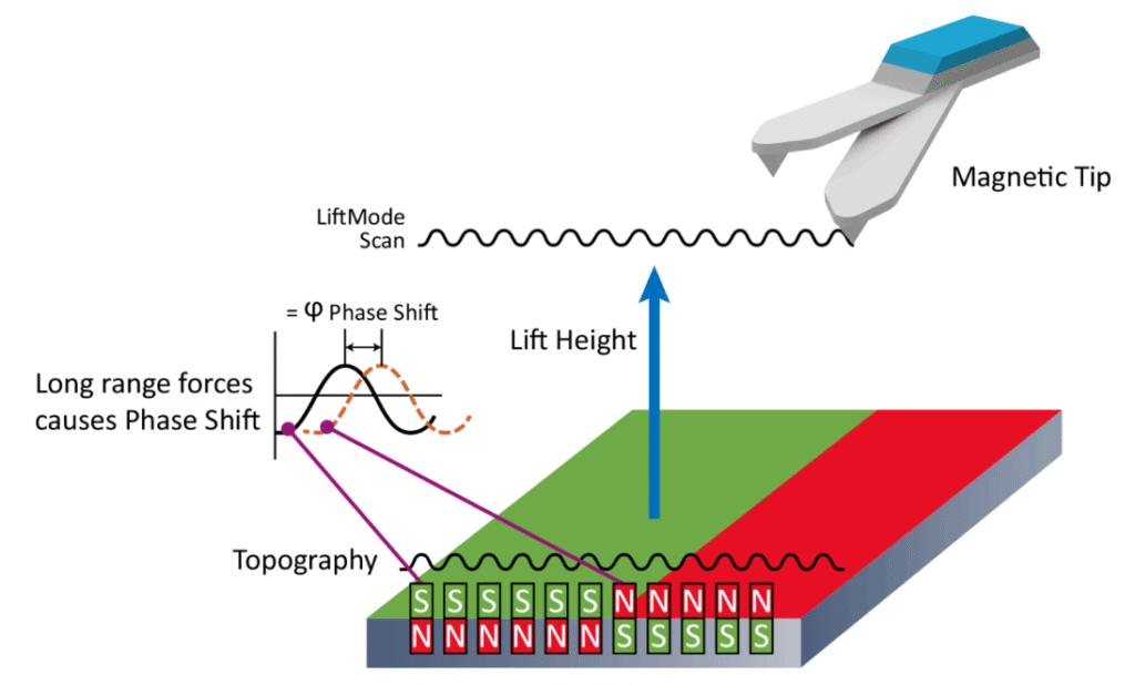
Magnetic Force Microscopy (MFM) enables the visualisation of stray magnetic fields at the sample surface by detecting the interaction between a magnetised cantilever tip and local magnetic domains. The imaging process involves two passes: the first to capture topography, and the second to trace the same line at a set lift height above the surface, recording phase shifts induced by long-range magnetic forces. Since these interactions decay slowly with distance, MFM is well-suited for resolving domain patterns at the nanoscale.
MFM signals are most strongly influenced by field gradients, and the resulting contrast—often observed in the phase signal—helps distinguish between regions of differing magnetic orientation or structure.
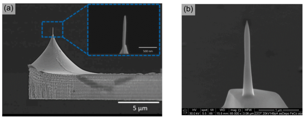
imaging. (b) Zoom-in of Co3Fe-based magnetic tip fabricated via FEBID
FusionScope’s Unique MFM Capabilities
The FusionScope platform enables a range of enhancements for magnetic imaging, distinguishing it from conventional MFM systems:
- Vacuum Operation:
FusionScope’s high-vacuum environment, required for SEM operation, also benefits MFM by improving signal-to-noise ratio. In vacuum, reduced damping leads to higher cantilever Q-factors, allowing more sensitive detection of magnetic interactions. A comparison presented in the application note shows nearly a 50% increase in phase contrast under vacuum compared to ambient conditions. - Self-Sensing Cantilevers:
FusionScope utilises piezoresistive self-sensing cantilevers, eliminating the need for external laser detection systems. This simplifies setup and reduces alignment complexity, particularly beneficial in a vacuum system. - Sharp FEBID-Fabricated Tips:
FusionScope uses Focused Electron Beam-Induced Deposition (FEBID) to fabricate sharp, high-aspect-ratio magnetic tips directly onto standard cantilevers. These tips, with radii of ~10 nm, enable high-resolution MFM imaging and improved lateral precision.
Discover the Quantum Design FusionScope
Correlative Imaging and Precision Tip Placement
A key feature of FusionScope is the Profile View, which allows the SEM column and MFM scan head to be tilted up to 80 degrees. This unique geometry offers researchers precise control over tip positioning, particularly important for small or complex sample features. By combining SEM imaging with real-time AFM/MFM scanning, users can reliably locate and target regions of interest, even those invisible to optical microscopy.
This correlative capability streamlines experiments involving lithographically defined samples or patterned nanostructures, improving workflow efficiency and measurement repeatability.

Application Examples from the Note
The application note illustrates the performance of FusionScope with two representative examples that highlight its capabilities in magnetic domain imaging:
- Permalloy Nanorod Arrays (Ni₈₁Fe₁₉):
Nanorod arrays fabricated by electron beam lithography serve as an ideal model system for exploring domain behavior and magnetic interactions at the nanoscale. FusionScope’s SEM imaging helped identify structures of interest, while MFM scans revealed localised magnetic signals at the nanorod vertices—where stray field gradients are strongest. This experiment underscores the utility of correlative SEM-MFM analysis for complex geometries. - FIB-Patterned Cobalt Thin Films:
In another example, a cobalt film was patterned using a focused ion beam (FIB) to create defined magnetic regions. SEM and AFM topography confirmed the pattern geometry, while MFM scans displayed clear cross-shaped domain structures. The lift phase images revealed magnetic features that were not visible in the continuous film, demonstrating how patterning alters domain configurations in a measurable way.
Conclusion: A Versatile Platform for Magnetic Imaging
The FusionScope® provides a powerful, integrated solution for researchers seeking to explore magnetic properties at the nanoscale. By combining high-vacuum SEM with AFM and MFM in a single instrument—and enhancing performance with FEBID tips and precision positioning tools—it enables detailed investigation of magnetic domains with clarity and control.
Whether studying magnetic thin films, nanostructures, or emerging materials with complex domain dynamics, FusionScope supports advanced scientific inquiry with a unique set of capabilities. The application note offers valuable insights into how this platform can be leveraged to uncover new phenomena and deepen our understanding of magnetism at the smallest scales.

To discuss your application…
Contact our Technical Sales Manager, Dr. Luke Nicholls, by email below or call (01372) 378822.


