There are two exciting new publications from our partners, NanoMEGAS. NanoMEGAS’ latest research on characterisation of Tang Dynasty gilded bronzes using Transmission Electron Diffraction (Phase and Orientation Mapping, precession assisted 3D ED, EDX), SEM (EBSD, EDX) and Synchrotron Methods has unveiled fascinating insights. And the paper in Nature Communications on 3D characterisation of colloidal assemblies in liquid is a great collaboration between Luis Liz-Marzán CIC biomaGUNE and NanoMEGAS.
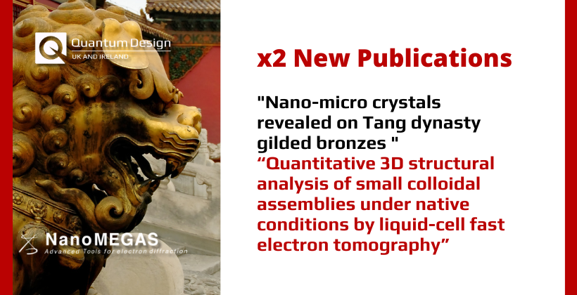
Nano-micro crystals revealed on Tang dynasty gilded bronzes using advanced TEM-SEM and synchrotron methods
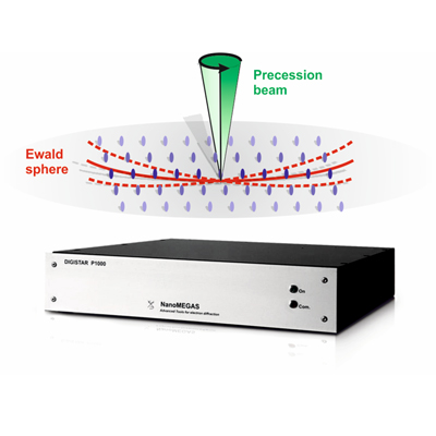
NanoMEGAS’ latest research on characterisation of Tang Dynasty gilded bronzes using Transmission Electron Diffraction (Phase and Orientation Mapping, precession assisted 3D ED, EDX), SEM (EBSD, EDX) and Synchrotron Methods has unveiled fascinating insights.
The work has been published in NanoScale, Royal Society of Chemistry Journal.
The study uncovered a copper-based core gilded with a thin gold layer, featuring an Au-Hg alloy in the headgear metal. The unique shaping and hammering techniques resulted in nanosized grains, revealing the artisans’ exceptional craftsmanship.
Abstract
Over the years, numerous gold and silver artifacts were excavated from tombs of the Tang Dynasty which gave evidence of the sophisticated metalworking techniques at that time. Few of the artifacts were thoroughly studied and their manufacturing processes were barely known. The present investigation concerns a metal headgear from a newly excavated female tomb in Xi’an of the Tang dynasty tomb (618-907 A.D.), using advanced techniques in a complementary way, especially performing a detailed analysis of the corrosion products and alloying processes. The combined state of the art methods and instrumentation used for the corrosion study included spectroscopy, diffraction, electron microscopy, synchrotron and their versions for specific measurements and sample preparation.
The investigated headgear metal consists of a copper-based core which was gilded by a thin gold layer, consisting of an Au-Hg alloy with a thin layer of about 400 nm. The technique used for shaping and the hammered embellishments led to the creation of nanosized grains on the side that would eventually be the interior of the headgear. It was gilded through the mercury-amalgam process, and the liquid diffusion caused the development of intermetallic compounds. This is the first recorded instance of these nano-scale and eutectic phases being observed on objects from an archaeological context. The crystallographic analysis offered valuable insights into the formation of needle-like malachite crystals growing on a layer of cuprite found on the surface of the corroded piece. The results highlight that the artisans utilised advanced methods in the creation of funerary items during the Tang Dynasty.
Quantitative 3D structural analysis of small colloidal assemblies under native conditions by liquid-cell fast electron tomography

Electron tomography has become a commonly used tool to investigate the three-dimensional (3D) structure of nanomaterials, including colloidal nano-particle assemblies. However, electron microscopy is typically done under high-vacuum conditions, requiring sample preparation for assemblies obtained by wet colloid chemistry methods. This involves solvent evaporation and deposition on a solid support, which consistently alters the nanoparticle organisation. Here, we suggest using electron tomography to study nano-particle assemblies in their original colloidal liquid environment.
To address the challenges related to electron tomography in liquid, we devise a method that combines fast data acquisition in a commercial liquid-cell with a dedicated alignment and reconstruction workflow. We present the advantages of this methodology in accurately characterising two different systems. 3D reconstructions of assemblies comprising polystyrene-capped Au nanoparticles encapsulated in polymeric shells reveal less compact and more distorted configurations for experiments performed in a liquid medium compared to their dried counterparts. A similar expansion can be observed in quantitative analysis of the surface-to-surface distances of self-assembled Au nanorods in water rather than in a vacuum, in agreement with bulk measurements. This study, therefore, emphasises the importance of developing high-resolution characterisation tools that preserve the native environment of colloidal nanostructures.

Interested in learning more about NanoMEGAS and your application?
Contact our Technical Director, Dr. Shayz Ikram, by email below or call (01372) 378822.