Coloration of a copper surface by nanostructuring with femtosecond laser pulses
Highlights
- Coloration of a copper by nanostructuring with femtosecond laser pulses was studied.
- Surface structures with low spatial frequency were formed.
- Surface topology on observed areas of different colours were investigated.
- SEM-images were taken in secondary electron and backscattered electron mode.
- Reasons for the colour difference are the surface topology and the oxidation degree.
Abstract
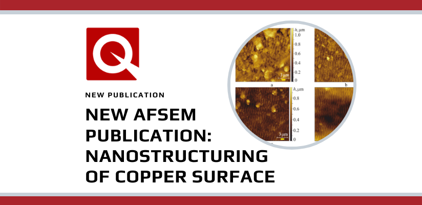
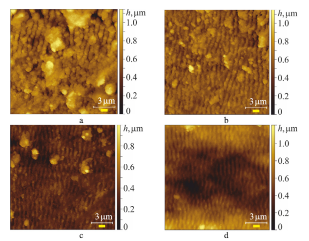
The coloration of a copper surface after treatment in air with femtosecond laser pulses at changing only the scanning direction has been studied. Surface structures with low spatial frequency, which were formed by a relative movement of the pulsed laser beam across the sample surface with an energy density below the ablation threshold, changed the sample colour. Four areas have been identified showing different colours such as blue-turquoise, orange, grey-green and red-violet. The possible influence of the surface topology on observed areas were investigated by atomic force microscopy. It was found that in a blue-turquoise area of the observed colour perception occurs the maximum average height of the relief and it is 1.7 times higher than in the red-violet area, while gray-green and orange areas have an average height between these values and are much closer to the values of the red-violet area. Scanning electron microscope images of the sample surface were taken in secondary and backscattered electron modes. It was revealed that the sample shows a combined microrelief consisting of low-spatial-frequency laser-induced periodic surface structures with an additional nanoroughness. It was determined that reasons for the colour difference of images are both the surface topology, as well as the degree of oxidation.
