


How PiFM Outperforms Conventional Techniques
Introduction
Coating the surface of acetaminophen particles with Al₂O₃ and ZnO via atomic layer deposition (ALD) has been shown to effectively modulate drug release rates [1]. The success of these approaches fundamentally depends on the uniform and conformal application of ALD coatings [2]. This, in turn, raises a critical question: how can we rigorously confirm the successful formation of an ultrathin layer? Addressing this question necessitates a technique capable of providing surface-sensitive chemical information with high spatial resolution.
Current methods
While surface-sensitive techniques such as TOF-SIMS can detect the presence of Al₂O₃, they are limited to identifying its existence rather than providing comprehensive information on its distribution or coverage across the particle surface. Merely establishing the presence of Al₂O₃ at isolated locations is insufficient for validating uniform ALD deposition, thereby excluding TOF-SIMS and similar methods from consideration for this application.
Techniques such as SEM/EDS and TEM-EDS may also be considered; however, the integration of their signals over significant sample thicknesses results in a lack of true surface sensitivity, especially for characterising ultrathin or single-molecule layers. In practice, SEM/EDS and TEM-EDS are far more likely to yield dominant drug substrate and elemental signals, with little to no sensitivity to the ALD overlayer.
ATR-FTIR is another candidate technique, utilising an evanescent wave generated at the interface of a high-refractive-index crystal to probe the sample. Figure 1 demonstrates the application of ATR-FTIR to Al₂O₃ ALD on acetaminophen. However, as evident from the spectra, ATR-FTIR lacks the required surface sensitivity for this application. The spectra of untreated acetaminophen and that of acetaminophen after 50 ALD cycles are essentially indistinguishable.

Unique PiFM approach
Clearly, the techniques discussed in the previous section do not have the spatial resolution or surface sensitivity to investigate ALD films. In contrast, photo-induced force microscopy (PiFM) offers a uniquely suitable solution. PiFM’s capability to fix the excitation laser at a specific wavenumber and raster scan the sample enables the collection of chemical maps with exceptional sub 5 nm spatial resolution. Once regions of interest are identified, single-point, surface-sensitive wavenumber sweeps can provide detailed chemical spectra from the very surface.
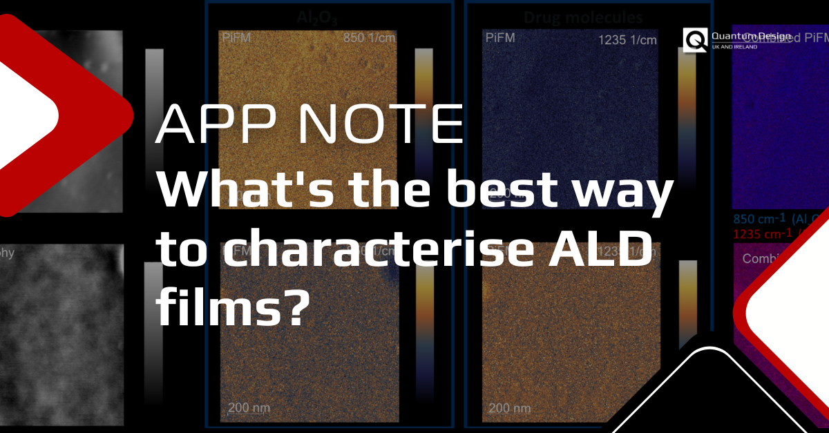
Figure 2 presents PiFM data for Al₂O₃ films on an unnamed drug molecule. Samples A and F possess unknown levels of Al₂O₃ ALD film on the drug particles. AFM topography images for both samples reveal surface roughness or aggregation features approximately 10 nm in height. Because the surfaces look so similar, the topograhy cannot provide any hints to determine if an ALD monolayer has been formed. Therefore, we can apply PiFM to gain chemical data on the sample. By tuning the PiF excitation laser to 850 cm−1 (the Al₂O₃ signature), PiFM imaging reveals a strong Al₂O₃ signal in sample F and a weak signal in sample A. Conversely, imaging at 1235 cm−1 (the drug IR signature) yields a strong drug signal in sample A and a weak signal in sample F. These results indicate that sample F is coated with a uniform ALD monolayer, whereas sample A exhibits none or very little ALD coverage. Therefore, PiFM allows us to gain localised chemical information about these surfaces, which is not something that any of the other analytical techniques considered could provide for this application.

In addition to the PiFM chemical maps, we can corroborate these findings using PiF-IR point spectra and comparing them to various references as shown in Figure 3. Each PiF-IR spectrum in Figure 3 is an average of 6 spectra acquired with a ~150 nm pitch. The FTIR control spectrum for Al₂O₃ (grey) displays no distinct peaks and a gradually increasing signal starting at 1100 cm−1. The control uncoated drug particle spectra, acquired via ATR FTIR (pink), shows the IR peaks associated with the drug molecule. The averaged PiF-IR spectrum taken on the uncoated drug particle (blue) shows peaks that agree well with the peaks from the pink reference spectrum on the uncoated drug material ATR FTIR spectrum. The PiF-IR spectrum taken on Sample A (green spectrum) exhibits features matching the uncoated drug particles, with no increase in peak strength below 1100 cm−1. This is consistent with the low signal intensity in the PiFM image at 850 cm−1 in Figure 2. Sample F’s PiF-IR spectrum (red) displays a pronounced Al₂O₃ signal, as indicated by the broad, rising baseline from 1100 cm−1. This is indicative of a more continuous Al2O3 layer, as supported by Figure 2. Notably, faint drug-related peaks are still observable in sample F, demonstrating PiFM’s ability to probe through ultrathin ALD films. Ultimately, these point spectra are consistent with Figure 2, strengthening the spotty and complete monolayer diagnoses we made using the single wavenumber PiFM images. Therefore, combining PiFM imaging with PiF-IR point spectroscopy allows detailed and accurate chemical characterisation at points of interest determined from topographical images.

In summary, PiFM stands out as the only technique capable of simultaneously delivering surface-sensitive, spatially resolved, and chemically specific data on ALD coatings. The integration of these three critical dimensions of information is essential for rigorously validating the uniformity and integrity of ALD surfaces. While the data presented in this application note used an unknown drug molecule as the substrate, a similar analysis can be performed for any combination of substrate and thin film (for example, inorganic or organic substrate covered with inorganic or organic film in any combination).
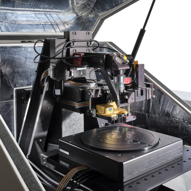
Molecular Vista “Vista 200” PiF microscope
For nano‑IR chemical analysis.
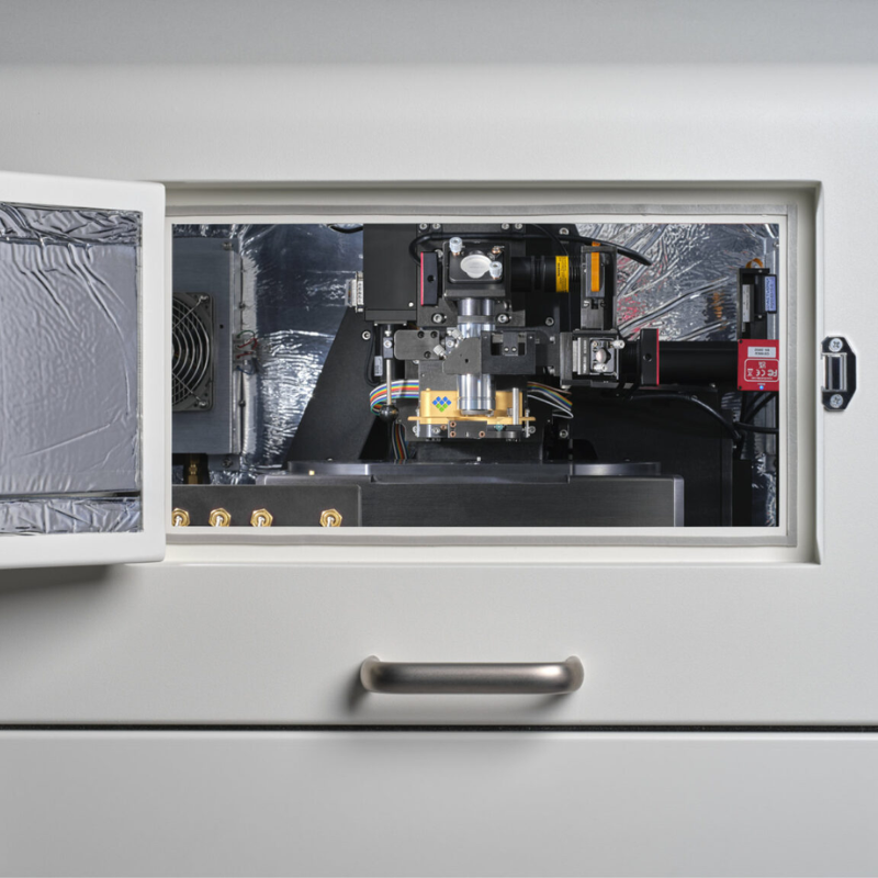
Molecular Vista “Vista 300” PiF microscope
Industry-leading automation

To discuss your application…
Get in touch with our Technical Director, Dr. Shayz Ikram, by email below or call (01372) 378822.
References
- Kääriäinen TO, Kemell M, Vehkamäki M, Kääriäinen ML, Correia A, Santos HA, Bimbo LM, Hirvonen J, Hoppu P, George SM, Cameron DC, Ritala M, Leskelä M. Surface modification of acetaminophen particles by atomic layer deposition. Int J Pharm. 2017 Jun 15;525(1):160-174. doi: 10.1016/j.ijpharm.2017.04.031. Epub 2017 Apr 18. PMID: 28432020.
- Boehler, C., Güder, F., Kücükbayrak, U. et al. A Simple Approach for Molecular Controlled Release based on Atomic Layer Deposition Hybridised Organic-Inorganic Layers. Sci Rep 6, 19574 (2016). https://doi.org/10.1038/srep19574


