- Models
- Downloads
- Applications
- Related Products
- Contact
- Back To Spectroscopy
- Back To Optics
- Back To Hyperspectral
- Back To Cameras
- Back To X-Ray
- Back To Light Measurement
- Back To Characterisation
- Back To Electron Microscopy
- Back To Magnetometry
- Back To Ellipsometers
- Back To Cryogenics
- Back To Lake Shore
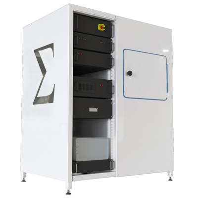
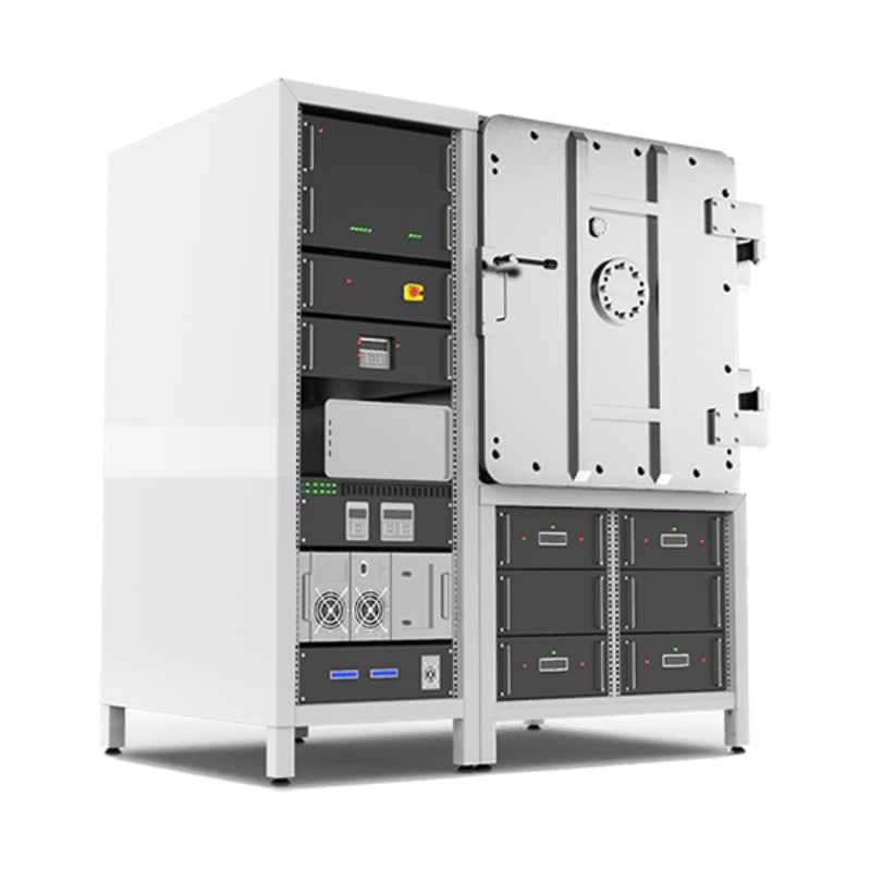
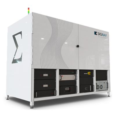
Sigray Attomap µ-XRF system
Highest Resolution XRF Microscope on the Market. Large Stage Travel and Enclosure
Sigray, Inc. develops advanced and completely new approaches to x-ray technology that formerly could only be found in synchrotron beamline experimental setups. The µ-XRF system from Sigray, AttoMap™ combines the resolution and the sensitivity from synchrotron XRF results and combines them into a laboratory-based instrument. Beside the AttoMap™ x-ray fluorescence system, Sigray is also offering proprietary x-ray optics and a new and breakthrough x-ray source named FAAST™.
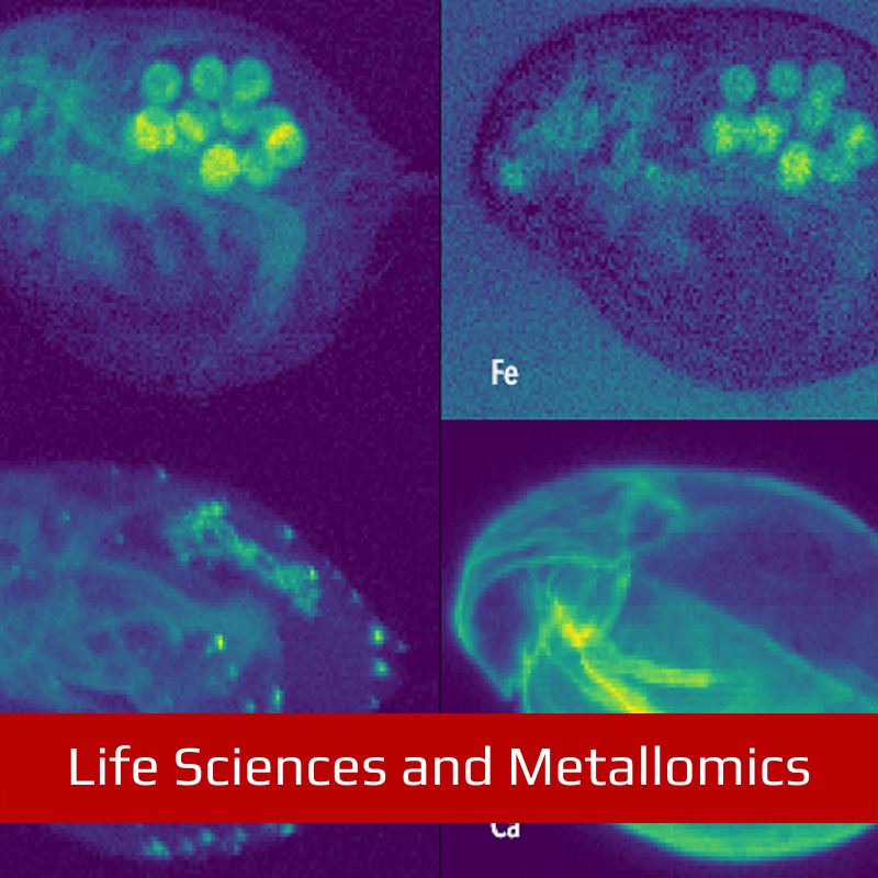
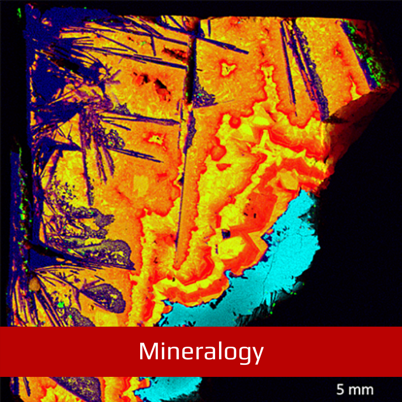
The powerful sensitivity and high resolution of the AttoMap produces synchrotron-quality elemental distribution mapping of trace elements for a wide range of research applications, spanning from the life and materials sciences to industrial use for pharmaceuticals, natural resources (oil and gas, mining) and semiconductor failure analysis. Click here for an amazing gallery of MicroXRF results
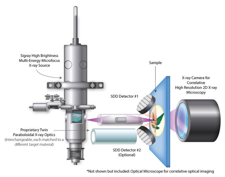
The AttoMap™ x-ray analytical microscope offers the highest resolution and the highest sensitivity one can find in a laboratory based microXRF system.
The AttoMap™ system can be used for transmission-based x-ray structural analysis as well as for fluorescence chemical mapping. The system has a chemical sensitivity of <1-10 ppm for trace element analysis and the measuring time is within 1 second.
The patented FAAST™ microfocus x-ray source (Fine Anode Array Source Technology) is based on a complete new x-ray source design. The x-ray target is made out of fine metal microstructures that are encapsulated in a diamond substrate. This complete new design was enabled due to recent developments in semiconductor processing techniques.
Models
Key Advantages
- Highest resolution laboratory microXRF Achieve down to single digit microns (3-5 µm) with high resolution optic
- Sub-ppm sensitivity Quantify down to sub parts per million (ppm) levels with Sigray’s flexible software packages
- Energy tunability Maximise throughput and sensitivity with up to 5 different incident x-ray spectra
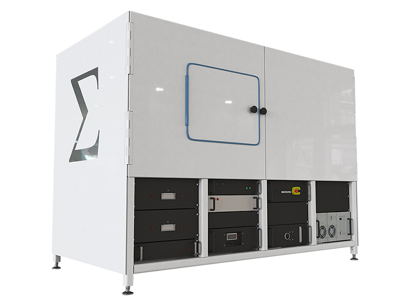
- Large Travel and Enclosure Enables unsupervised overnight scans. Also provides opportunity to integrate correlative techniques such as Raman Spectroscopy
System Features
- FAAST™ microfocus X-ray source with 50x higher brightness than sources used in existing non-synchrotron microXRFs due to innovative X-ray target material design and provides up to 5 different spectra in a single source
- X-ray mirror lens unique design collects 10x more fluorescence X-rays versus conventional designs
- 500x higher throughput versus existing synchrotron and non-synchrotron microXRFs
- Optional dual energy source for maximum flexibility
- Quantitative Analysis with standards-based or standardless approaches with wide range of flexible and intuitive software routines, from mineralogy to semiconductor-focused wafer pattern navigation, flexible and customisable Jupyter notebooks, and fundamental parameters analysis for weight percentages
- >1000X sensitivity of SEM-EDS for simultaneously acquiring a range of elements with the ability to measure thin films and dopants at sub-angstrom sensitivity and nanometer resolution.
- Chemical imaging at <8um and down to 5ms/point, with trace element detection in seconds
Key Advantages
All the benefits of the AttoMap-200, plus, advances exclusive to the AttoMap-310, including:
- Achieves shallow angle imaging for thin samples (e.g., biological) and/or diffraction removal AttoMap-310 features a goniometer to enable normal to shallow angle of incidence imaging
- Light element detection Analyse down to trace-level organics within the AttoMap’s high vacuum chamber
System Features
- Patented high brightness x-ray source with 50X brightness over those used in other leading microXRF systems and provides up to 5 different spectra in a single source
- Mirror Lens x-ray optics with major advantages over conventional polycapillary microXRF systems
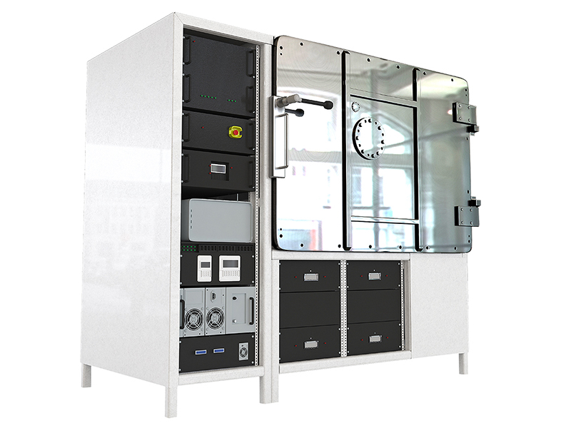
- Goniometer stage for variable angle imaging (3 to 90 degrees)
- Vacuum enclosure that achieves down to <10^-4 Torr
- Wide range of flexible and intuitive software routines, from mineralogy to semiconductor-focused wafer pattern navigation, flexible and customisable Jupyter notebooks, and fundamental parameters analysis for weight percentages


