- Features
- Options
- Specifications
- Videos
- Downloads
- Applications
- Related Products
- Back To Spectroscopy
- Back To Optics
- Back To Hyperspectral
- Back To Cameras
- Back To X-Ray
- Back To Light Measurement
- Back To Characterisation
- Back To Electron Microscopy
- Back To Magnetometry
- Back To Ellipsometers
- Back To Cryogenics
- Back To Lake Shore
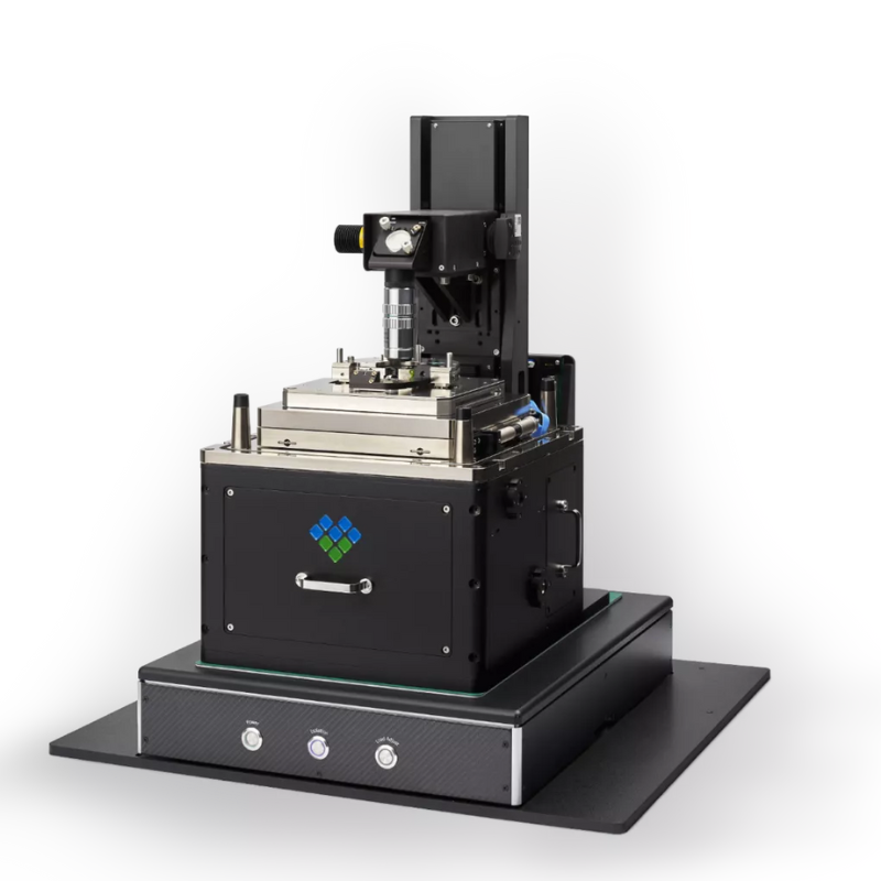
Molecular Vista “Vista One” PiF Microscope
Nano IR Microscope and Spectrometer
The original PiF microscope for nano‑IR chemical analysis
Vista One makes nanoscale chemical maps and point spectra with more detail than FTIR or nano-FTIR. Characterise at the highest resolution.
Offering the highest resolution nanoscale chemical analysis instruments available.
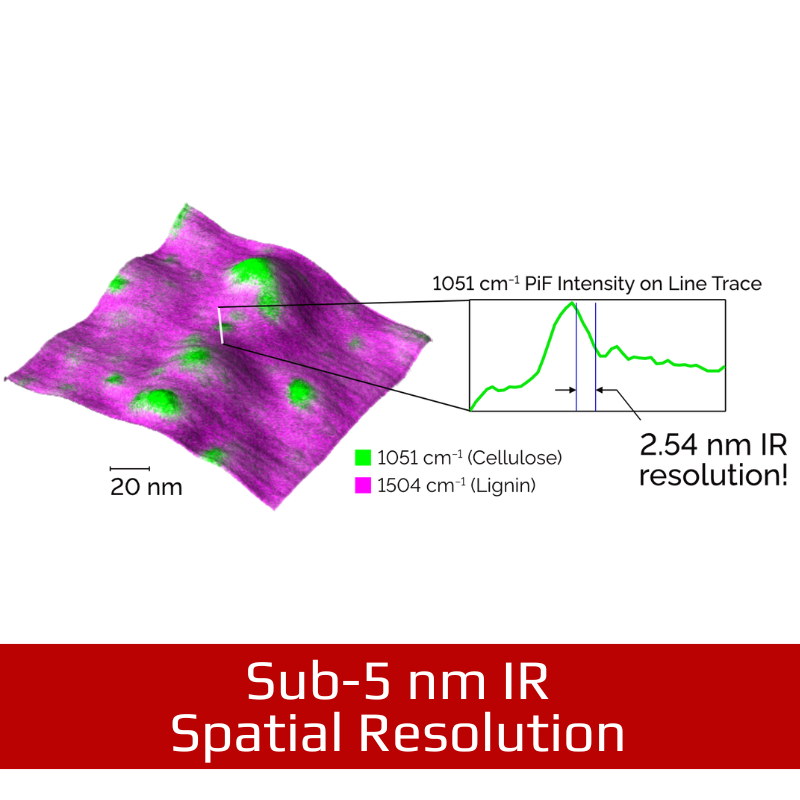
- Sub-5 nm IR spatial resolution
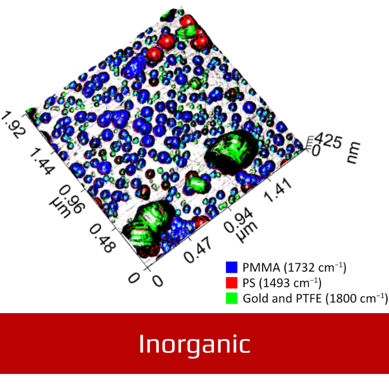
- Unique chemical identification capability
Complete results immediately
Designed for high throughput measurement
- Tunable IR lasers can sweep a full PiF‑IR spectrum in as little as 100 ms.
- Use existing FTIR spectra to identify materials.
- Fixed-wavelength PiFM images can be acquired in minutes with a range of up to 80 µm in X and Y.
- Optical alignments are maintained even when changing samples.
- hyPIR™ imaging and our automated principle component analysis tools provide complete image and spectral data‑sets with minimal effort.
Features
-
Engineered for precision
- Capacitive sensors + optical encoders – The motorised stage features 6 mm travel and optical encoders for precision control. The capacitive sensors in the AFM scanner ensure linear scans with a ~100 pm RMS precision.
- Dual Z – Image samples faster without introducing artifacts. The dual Z‑piezo scanner system provides accurate scans with a large 12 µm vertical range.
-
Proper optics integration
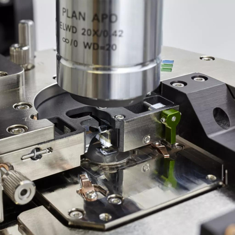
- Built to handle any light – A 3D-actuated parabolic mirror focuses excitation light onto the side of the tip no matter what wavelength of light is used.
- Fast cantilever alignment – The cantilever alignment chip ensures that optical alignments are maintained for both the AFM feedback and near-field lasers during cantilever changes.
-
Accessible and stable AFM head
- Invar construction – Both the head and scanner are made of invar for the best thermal stability possible.
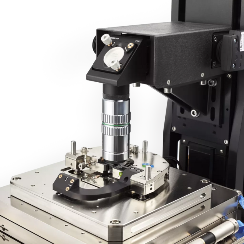
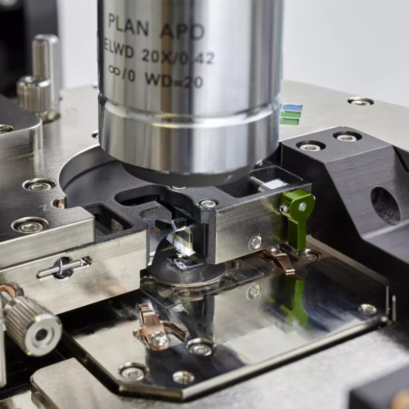
- Low profile stability – The low-profile AFM head allows us to use top objective lenses with high numerical apertures. This provides an excellent optical view of the sample. The head also features the most stable mount configuration for low drift and acoustic stability.
-
Versatile optical pathways
- Bottom optics* – Nano-precision independent scanning allows moving the excitation source focus spot in 3D space.
- Many optical views – Access the top, bottom, or side of the AFM tip via the top objective, bottom optics, and parabolic mirror. Configure up to 6 lasers for PiFM and PiF-IR.
-
Customisable environments
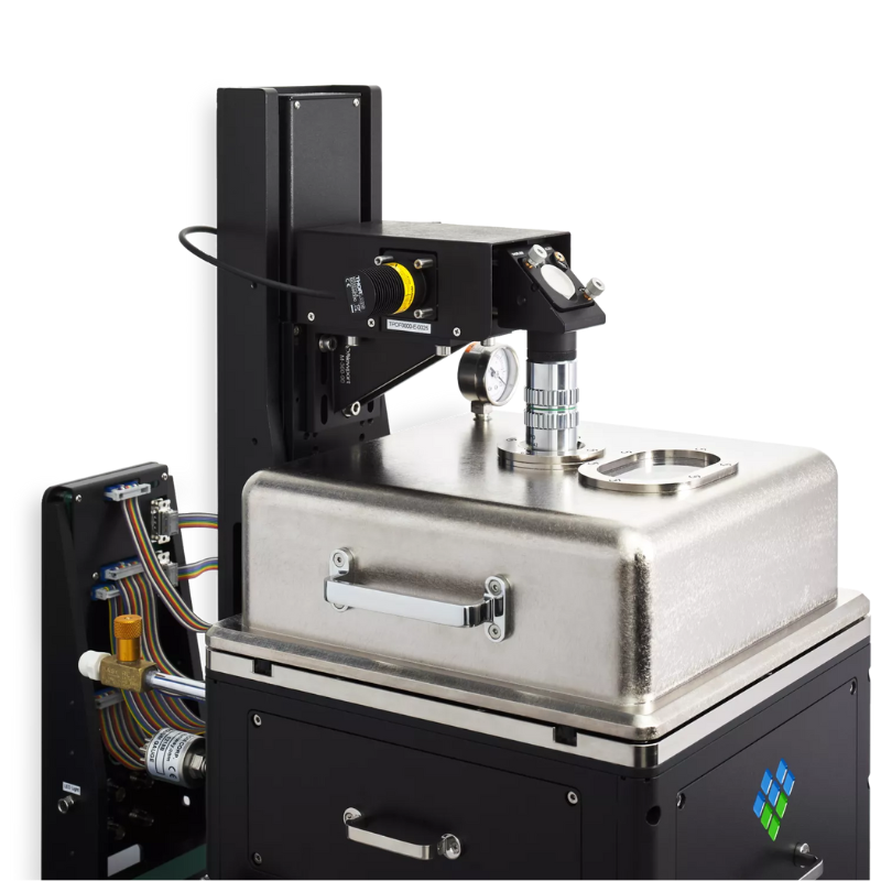
- Optional environmental cover – The optional environmental/vacuum cover allows customisation of the entire microscope environment. Work under partial pressure with any gas, pump down to vacuum, or control the humidity.
-
Work in your element
- No need to whisper – Install Vista One anywhere you want, and the combination of acoustic enclosure, active isolation table, and external cable frame ensure the AFM tip stays quiet.†
- Thermal isolation – The Acoustic Chamber features a temperature control unit to keep the entire system stable within ±0.1 ºC. *
-
Versatile and customisable
- Any optical technique – Vista One is Molecular Vista’s original PiF microscope designed to handle any optical experiment. It also supports sSNOM, confocal Raman, and many custom designed experiments.
- Customise any optic – Molecular Vista uses standard optical components from ThorLabs and other suppliers. This means that you aren’t tied into a proprietary system and your Vista One will be infinitely configurable by nature.

Specifications
| Beam Deflection AFM Head (AFM-BD) | Body Material: Invar for excellent thermal stability |
| Body Profile: 12 mm thick to allow short working distance top objective lens | |
| AC Detector Noise: <50 fm/root Hz above 100 KHz | |
| Detector Bandwidth: 6 MHz | |
| Cantilever Deflection Sensor Laser: 904 nm with finely adjustable beam steering and focus | |
| Manual Translation Stage: 3mm movement in X and Y for coarse tip alignment to focus point of optional bottom objective lens | |
| Fast-Z Module: 1 um z-piezo on cantilever mount serves as the Fast-Z element of high-speed Dual-Z feedback system | |
| Operational Environment: Ambient air, optional open liquid cell, or vacuum/partial pressure with optional environmental chamber cover | |
| Excitation Optics: Broadband (400 nm – 20 um) integrated parabolic mirror with 3D piezo-motor stage for tip-sample illumination for PiFM and reflection-mode s-SNOM | |
| Forward Facing Tuning Fork AFM Head (AFM-FFTF) | Body Material: Invar for excellent thermal stability |
| TF Operation: Tapping-mode | |
| Manual Translation Stage: 3 mm movement in X and Y for coarse tip alignment to external laser or focus point of bottom objective lens (for tip-enhanced spectroscopy) | |
| Integrated Tip Scanner: X-Y flexure scanner built into head with 12 um x 12 um range for scanning the tuning fork | |
| Fast-Z Module: 1 um z-piezo on cantilever mount serves as the Fast-Z element of high speed Dual-Z feedback system | |
| Main Body Frame | Versatile frame architecture provides for multiple optical pathways to the tip-sample interface. |
| Inverted Objective Lens (optional): 100X 1.4NA Oil immersion lens or 60X 0.9A conventional lens forms the basis of a custom-designed inverted optical microscope for bottom viewing and illumination of the tip-sample interface. Optional broadband reflective lens available for wideband IR illumination. | |
| Tip Alignment Mechanism: Piezo-driven XYZ stage (12 um for XY and 100 um for Z) for the inverted objective lens for precise alignment of the focus spot onto the tip | |
| Top Objective Lens: 20X, 0.42NA 20 mm working distance standard; shorter working distance (down to 13 mm) also supported for higher NA options | |
| Top Objective Lens Focus: Motorized with stored focus position for fast return after tip or sample change | |
| CCD Camera: Concurrent top and inverted views ; digital zoom, pan, and capture | |
| Tip-Sample Approach: Automated engagement | |
| Sample Stage: Motorized precision stage with 6 mm x 6 mm travel range | |
| Maximum Sample Size: 25 mm x 25 mm x 5 mm | |
| System Noise: <90 pm RMS (dependent on environment) | |
| Optical Configuration: Multiple optical pathways to bottom objective and side parabolic mirror provided; pathways are based on industry standard 1” cage system to allow user customization and expansion. | |
| Sample Scanner: XYZ flexure stage scanner with 80 um x 80 um x 15 um scanning range (closed loop); Z sample scanner serves as the slow Z component of high speed Dual-Z feedback system. Built-in capacitive sensors provide closed-loop scanning control for X and Y for superb linearity and accuracy; optional Z capacitive sensor available. | |
| Scanner Material: Invar for excellent thermal stability | |
| Scanner Sensor Noise: 0.15 nm RMS for X and Y with 0.08 nm RMS achievable with software controlled reduced scan range (20 um x 20 um) | |
| High-Speed Electronics | FPGA-based control electronics has a section dedicated for high speed scanning probe microscopy. |
| PiFM & Optics Electronics | TTL Signal Generator: Two flexible TTL signal generators (with 160 MHz sampling rate) with adjustable duty cycle and DC offset for direct current modulation of laser diodes or for input to Bragg cells |
| Flexible Lock-in Referencing: Lock-in amplifiers can be phase locked to any other lock-in or at any calculated frequencies from the other lock-ins | |
| Digital Counter Input: Input for avalanche photodiode or photomultiplier for low-light detection | |
| Computer | Mounted in a 19″ rack. Minimum configuration includes i7 based Quad Core, 8GB RAM, 256GB SSD and 2000GB HD combination, 26″ or dual monitor support , 8×USB ports, Windows 10 64bit Professional |
| VistaScan Image Acquisition Software | Supported modes/features include: |
| Contact and AC AFM | |
| STM and PLL feedback (for high Q sensors such as tuning-fork) | |
| Ultrafast Dual-Z feedback | |
| Q-control | |
| Bi-modal force gradient imaging for linear and non-linear PiFM | |
| Sideband force gradient imaging (for KPFM via electric force gradient detection) | |
| Concurrent acquisition of 26 channels in Dual-Z configuration and 40 channels in Slow-Z configuration | |
| Concurrent acquisition of 4 channels for each spectroscopy mode which may include | |
| vs gap distance | |
| vs bias with and without feedback | |
| step response to voltage response with and without feedback | |
| SurfaceWorks Image Analysis Software | Powerful and intuitive software. Features include: |
| Shape and histogram-based masks | |
| Functions and analysis (flattening, FFT filtering, line & region analysis, 3D rendering, palettes, etc) applied to an image saved as a file property along with the raw data file | |
| Copy/Paste file property to apply same functions and analysis to other image file(s) | |
| Preview feature for most functions | |
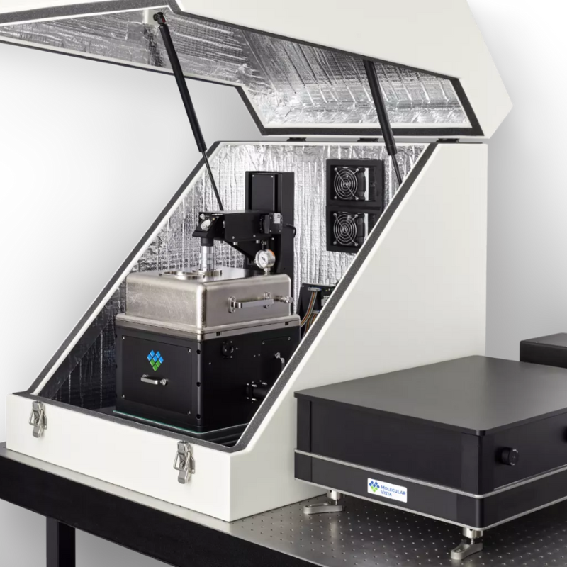
| Acoustic Enclosure | Optional acoustic enclosure (30W×30D×27H in3). Available with or without temperature control. |
| Comes with ports for cables and optical access. |
VIDEOS
Downloads
Supplier Info
APPLICATIONS
Sub-5 nm IR spatial resolution
PiFM chemical mapping provides unbeatable lateral IR resolution for analysing surface chemistry. An elementary cellulose fibril is embedded in the lignin matrix of a cell wall from spruce wood. Scan dimensions: 150 nm × 150 nm × 10.5 nm.
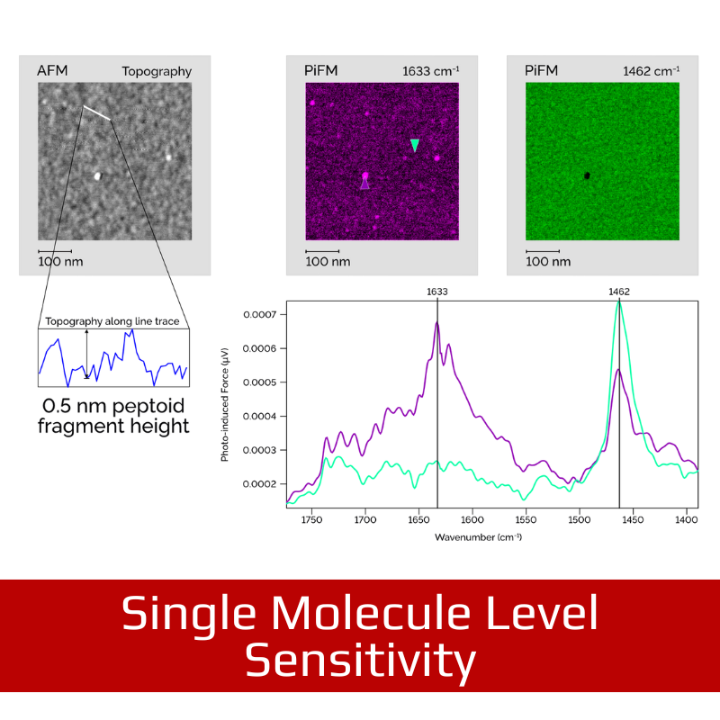
Single-molecule level sensitivity
PiF-IR PiF-IR spectra (magenta and green) show the chemical differences on and off these peptoid nano-sheet fragments. PiFM PiFM chemical maps are able to detect peptoid fragments that are only 0.5 nm taller than the surrounding material! Scan dimensions: 500 nm × 500 nm × 1.7 nm.


