Empowering Discovery at the Frontiers of Life Science
At the intersection of technology and biology, innovation is accelerating faster than ever. From molecular imaging to advanced spectroscopy, high-performance instrumentation is redefining what scientists can see, measure, and achieve. Quantum Design UK and Ireland proudly support this transformation—providing state-of-the-art tools that enable researchers to explore the deepest complexities of life and health.
To celebrate this spirit of innovation, we’re excited to introduce the inaugural issue of our new publication, NextGen BioTools—a free magazine dedicated to the forefront of high-tech instrumentation for life science and biomedical research.
Within its pages, you’ll discover how pioneering researchers and organisations are using advanced technologies to push scientific boundaries. Highlights include an exclusive interview with Professor Hans Arwin, a world authority on spectroscopic ellipsometry, insights into the rapidly evolving field of correlative microscopy, and a behind-the-scenes look at how Boston Scientific leverages next-generation tools to drive biomedical innovation.
From precision instruments to powerful new methodologies, NextGen BioTools is your window into the technologies shaping the future of biological discovery.
Join us in exploring the next generation of scientific innovation—download your free issue today.
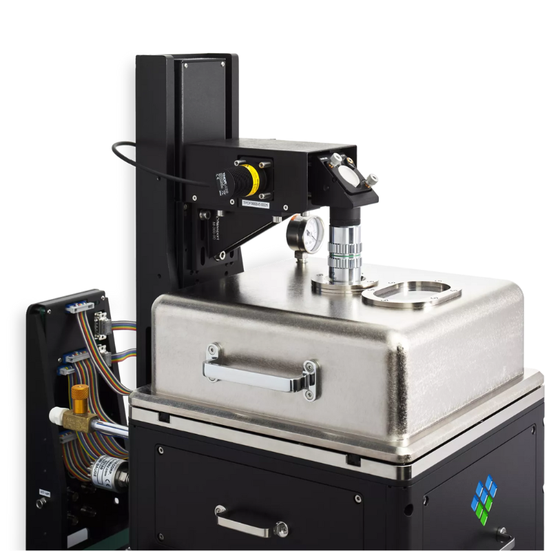
Easy to use Correlative AFM with SEM Microscopy Platform
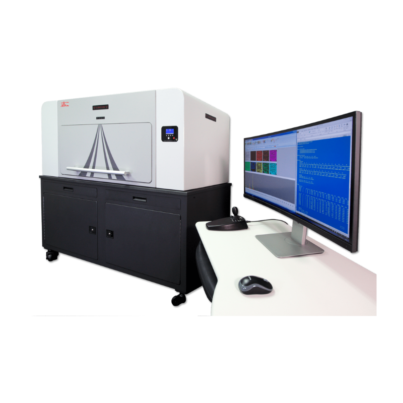
Micro X-ray Fluorescence (μXRF) Hyperspectral Imaging Elemental Analyser
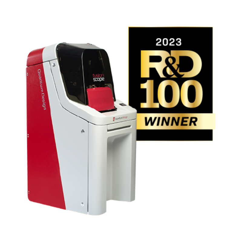
Nano IR Microscope and Spectrometer


IXRF Systems’ ATLAS M benchtop microEDXRF (micro XRF) spectrometer is the latest general purpose micro spot energy dispersive X-ray fluorescence (EDXRF) spectrometer microscope for the measurement and mapping of elements from sodium (Na) through uranium (U). Designed to image and analyse a wide variety of sample types, ATLAS leads the industry in virtually every major specification category from the most powerful software and the highest detector active area, to our superior perpendicular (normal) X-ray tube geometry geometry and smallest micro spot.
Micro X-ray Fluorescence (μXRF) Hyperspectral Imaging Elemental Analyser
Nanoscale Chemical Imaging of Soft Biological Tissues
Exploring the capability of PiFM on fixed human skin samples that were penetrated by topically applied dexamethasone and show that there is similarly a good correlation between XAS microscopy and PiFM, with PiFM offering much higher spatial resolution.
A cross-section of prepared skin slices of 300 nm thickness was deposited on a Si3N4 window for XAS microscopy; the same area was then subsequently analysed by PiFM. Given the FTIR spectrum of dexamethasone (Fig. 1a), PiFM images were acquired at 1708 cm−1 and 1559 cm−1 to highlight dexamethasone molecules and corneocytes (via amide II band), respectively; amide I band was avoided since dexamethasone has a strong absorption at around 1660 cm−1.
ALL APPLICATIONS

Defence and Security

Aerospace

Quantum Science and Technology

Space and Astronomy

Semiconductors

Agriculture
Non-Destructive Testing