- Features
- Models
- Specifications
- Downloads
- Applications
- Related Products
- Contact
- Back To Spectroscopy
- Back To Optics
- Back To Hyperspectral
- Back To Cameras
- Back To X-Ray
- Back To Light Measurement
- Back To Characterisation
- Back To Electron Microscopy
- Back To Magnetometry
- Back To Ellipsometers
- Back To Cryogenics
- Back To Lake Shore
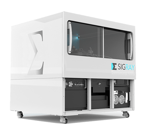
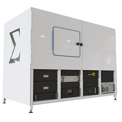
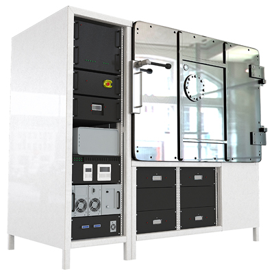
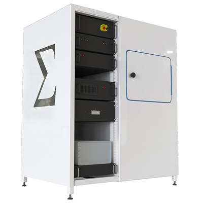
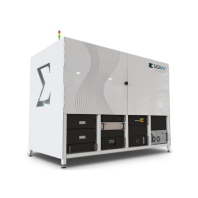
Sigray QuantumLeap-H2000 X-Ray Absorption Spectroscopy (XAS) System
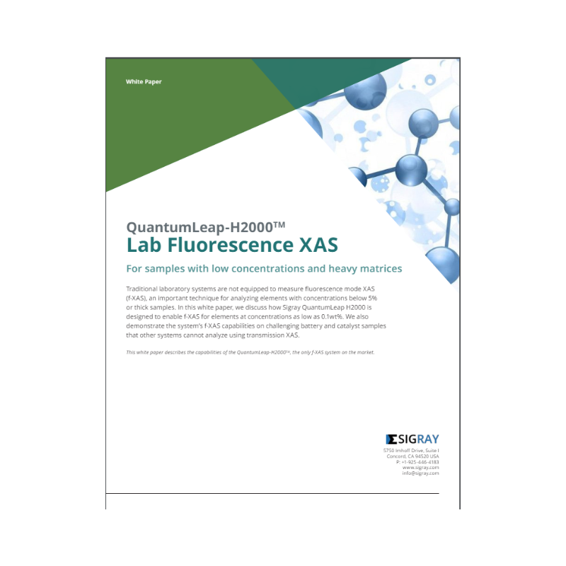
First laboratory XAS system with both transmission and fluorescence mode XAS
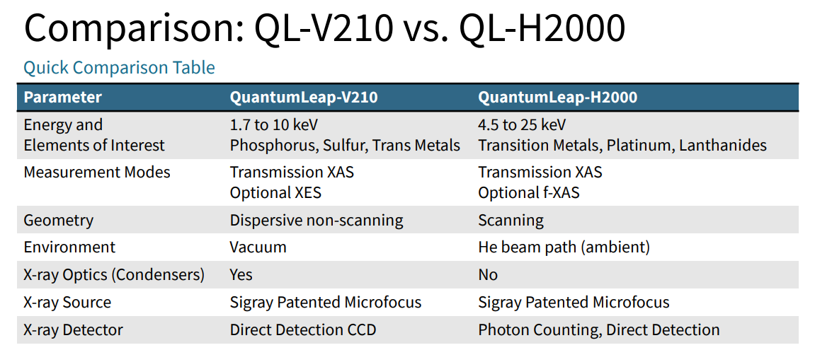
The first commercial laboratory-based Hybrid XAS System offering both transmission and fluorescence XAS modes, in scanning geometry. System is in ambient conditions with the line focus spots reaching 50um in short dimension and 1-2mm in the long dimension.
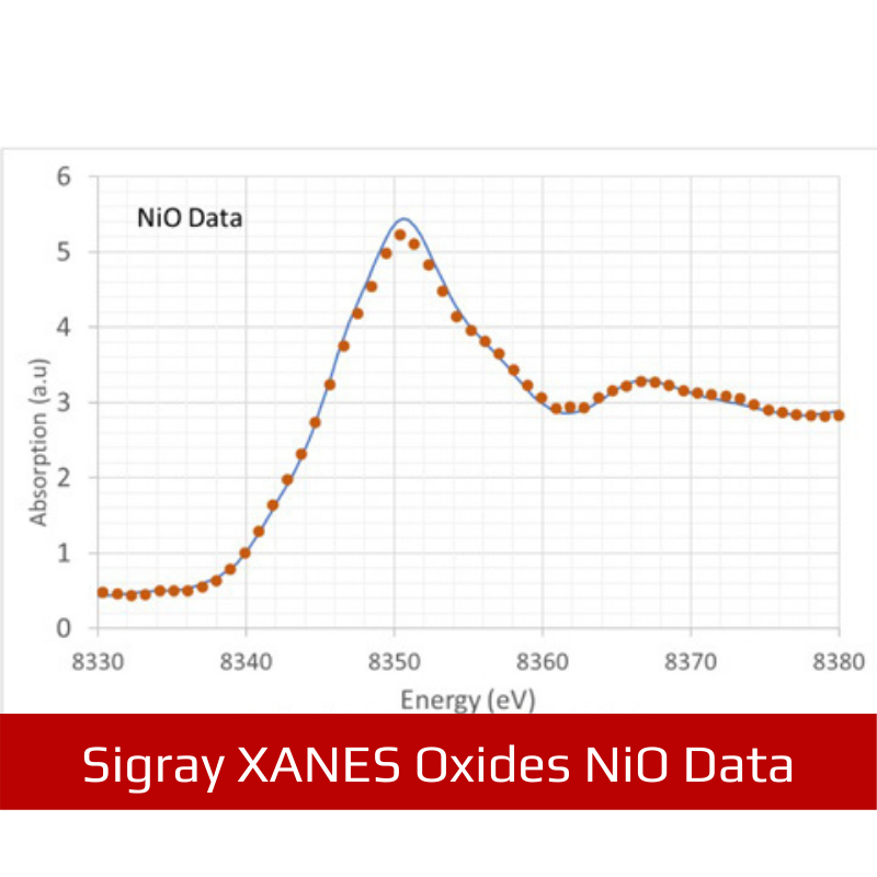
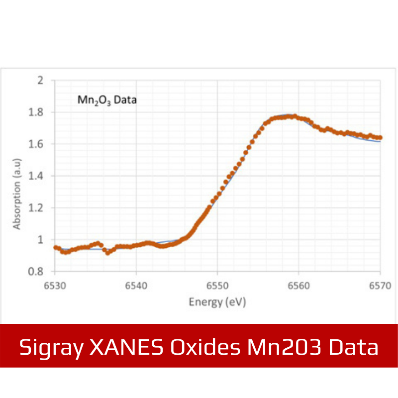
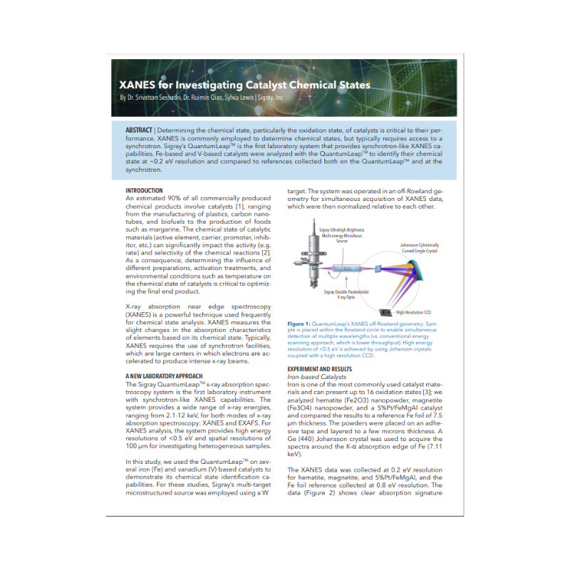
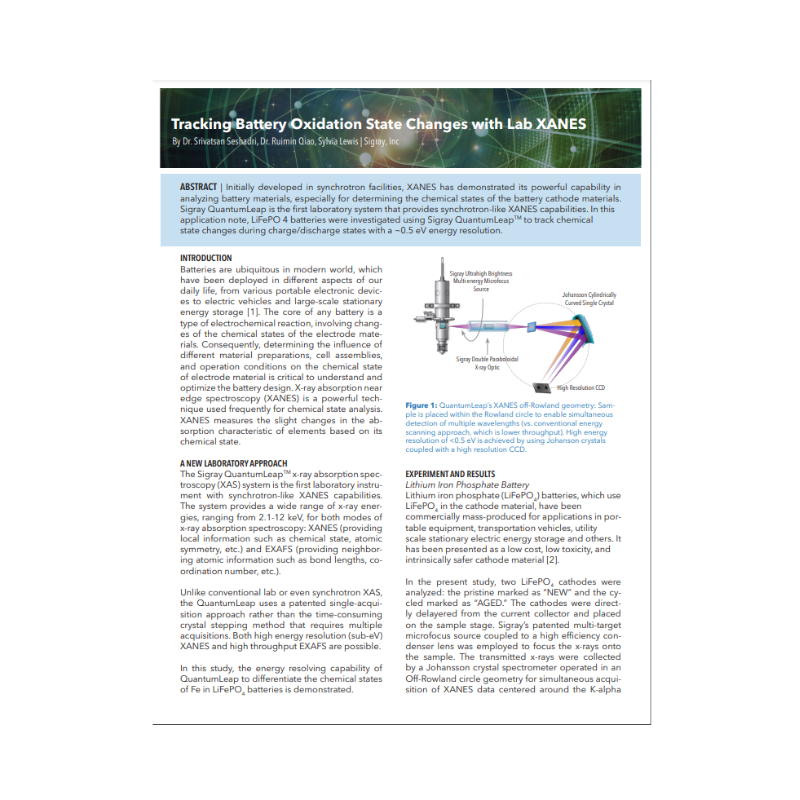
Specialised for achieving synchrotron-grade XAS, in both transmission and fluorescence modes, in ambient-based laboratory conditions to provide chemical state and electronic structure information
Synchrotron-like Performance in a Laboratory XAS System
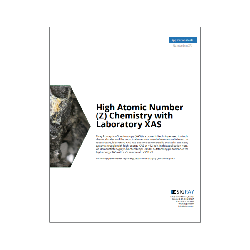
Compared to conventional high Bragg angle laboratory XAS systems, the Quantumleap H2000 has significant advantages including:
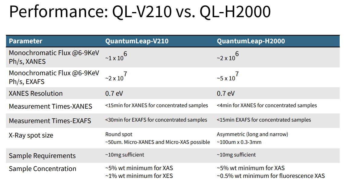
- Optimised throughput of 5X flux for XANES and 20X flux for EXAFS
- Only 4 crystal analysers are needed to cover the complete energy range of 4 to 20 keV (whereas multiple crystals may be required for a single 1 keV XAS spectrum in high Bragg angle systems). The small number enables the system to include all crystals on a software selectable robotic stage.
- Constant spot profile unlike for high Bragg angle XAS systems, the spot size changes significantly for every crystal rotation which places restrictions on sample uniformity.
- Flux for operation in fluorescence-mode
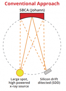
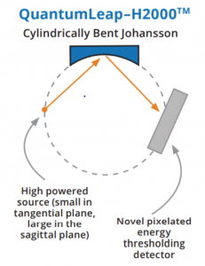
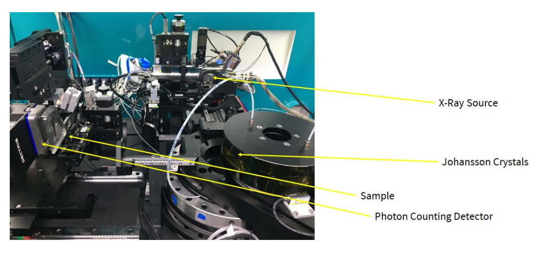
Conventional laboratory XAS systems [left] use large spot sized, high powered x-ray sources, a Johann spherically bent crystal analyser (SBCA), and a silicon drift detector; such designs operate at high Bragg angles on a large Rowland circle. Sigray’s QuantumLeap-H2000 uses a high powered x-ray source that has a small dimension along the tangential direction and large dimension in the sagittal direction (which in this illustration, is into the paper). The source, along a Johansson crystal and a novel pixelated energy thresholding detector, enables advantageous operation at low Bragg angles.
XAS GALLERY LINKS:
FEATURES
Specialised for achieving synchrotron-grade XAS, in both transmission and fluorescence modes, in ambient-based laboratory conditions to provide chemical state and electronic structure information
- Patented design enabling acquisition at low Bragg angles (e.g. 15 to 30 degrees) through use of a Johansson crystal in combination with a photon counting detector. Therefore, stitching data is not needed (which results in artifacts and slower data acquisition).
- Patented X-ray source with outstanding brightness and a multi-target design for ideal spectral output
- Patented acquisition approach to optimize XAS spectra at highest throughput, achieving down to 0.5eV energy resolution
Comparison
SPECIFICATION
| Parameter | Specification | |
|---|---|---|
| Overall | Energy Coverage | 4.5 to 25 keV |
| XAS Acquisition | Transmission mode Fluorescence mode |
|
| Energy Resolution | Sub-eV in XANES 5-10 eV in EXAFS (Note that you can also use XANES mode to acquire high resolution EXAFS) |
|
| Beam Path | Helium flight path | |
| Focus at Sample | Line focus: 30-100 μm in one direction; ~300 um – 3mm in other direction | |
| Source | Type | Sigray patented ultrahigh brightness sealed microfocus source |
| Target(s) | W and Mo standard. Others available upon request. |
|
| Power | Voltage | 300W | 20-50 kVp | |
| X-ray Crystals | Type | 2 Johansson single crystals 1 mosaic crystal |
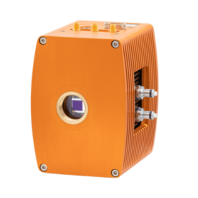
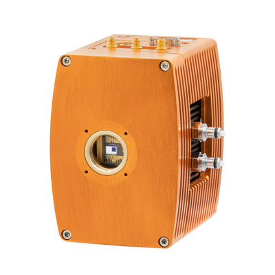
| X-ray Detector(s) | Type(s) | Spatially resolving (pixelated detector) for transmission XAS Silicon drift detector (SDD) for fluorescence XAS |
| Count Rate | 10^8 x-rays/s for photon counting detector 500k cps for SDD |
|
| Dimensions | Footprint | 53″ W x 77″ H x 66″ D |


