- Features
- Models
- Specifications
- Videos
- Applications
- Related Products
- Contact
- Back To Spectroscopy
- Back To Optics
- Back To Hyperspectral
- Back To Cameras
- Back To X-Ray
- Back To Light Measurement
- Back To Characterisation
- Back To Electron Microscopy
- Back To Magnetometry
- Back To Ellipsometers
- Back To Cryogenics
- Back To Lake Shore
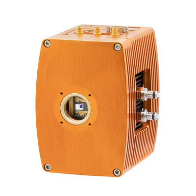
Sigray TriLambdaXRM-30 3D X-Ray Microscope
Nano X-ray Microscope
Spatial resolution to 35nm
Up to 3 X-ray Energies in Single System
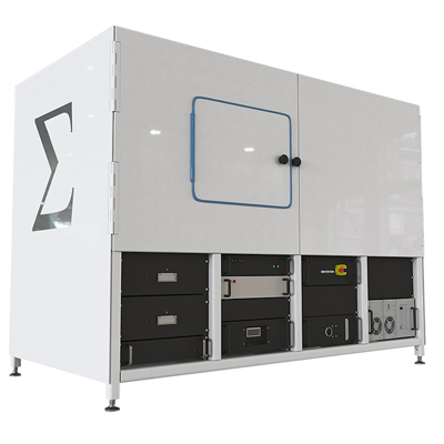
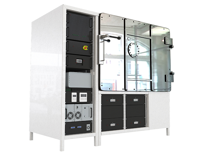
The Trilambda XRM-30 system: this NanoCT with patented X-ray and optics is designed for ultra-high spatial resolution (35nm) imaging with up to 3 X-ray energies in a single system.
FEATURES
- 3D nano x-ray microscope (nanoXRM) with 35nm resolution
- Highest resolution system on the market. Enabled by x-ray optics.
- Multiple x-ray illumination energies
- Up to three energies selected in a single system for optimised imaging for a broad range of samples (polymers to metals)
- Dual energy nano tomography for quantitative information
- Density and improved contrast can be obtained by taking tomographies at two different energies
HIGHLIGHTS
The TriLambda nanoXRM achieves over an order of magnitude in superior resolution compared to lensless x-ray microscopes such as the PrismaXRM, which do not use x-ray optics. Such resolution is achieved through the use of Sigray’s proprietary double paraboloidal x-ray optics and diffractive focusing zone plate optics. PrismaXRM and TriLambdaXRM provide natural multi-scale complements to each other; PrismaXRM provides submicron imaging of larger samples, while the TriLambda provides 35-100nm resolution imaging of smaller samples.
Image: Outstanding 35-50nm resolutions can be achieved for x-ray energies ranging from 2.7 keV to 8.04 keV.
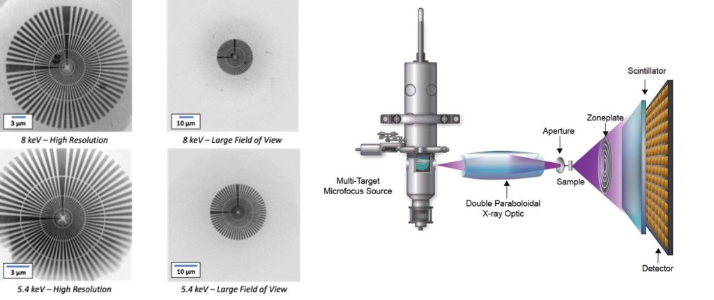
Up to three x-ray energies are included in the TriLambda system, providing optimal illumination for the wide range of samples seen in a busy central laboratory. X-ray contrast varies significantly for each x-ray energy, impacting both the time to acquire data and more importantly, the visibility of structures. For example, 5.4 keV is optimal for geological samples (SiO2) but poor for samples with metal in them. For polymers and cells, soft x-ray energies (2.7 keV) provide profound advantages over 8 keV.

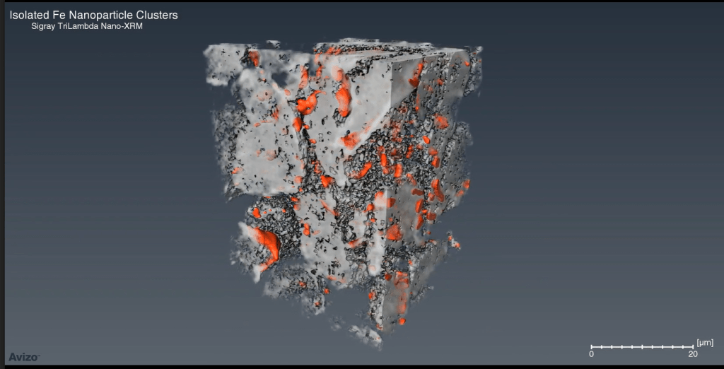
Interpreting contrast measurements and CT numbers can be a difficult task when only a single X-ray energy is used for imaging. This is due to the presence of sub-resolution features, such as nanoporosity or nano-scale alloying elements that change the absorption of a sample and lead to errors in image segmentation and inaccurate quantification of the atomic number of a material. By imaging at two different x-ray illumination energies (dual energy), TriLambda removes such errors.
Image: 3D surface rendering of segmented Fe NPs (orange) in relation to the graphite clusters acquired with dual contrast
TriLambda’s x-ray source features a patented design in which multiple target materials are in optimal thermal contact with diamond, which has excellent thermal conductivity properties. The rapid cooling of diamond enables higher power loading on the x-ray source to produce an intense beams of x-rays. This thermal benefit enables more materials to be used as x-ray source target materials, each of which produce strong characteristic x-rays of a specific energy. Up to 3 target materials can be customized for the TriLambda-30 source, allowing software selection of the optimal spectra for your sample.
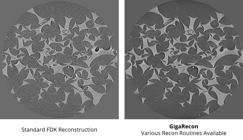
GigaRecon | Tomographic reconstruction software that pairs the fastest reconstruction times with an unmatched suite of features for achieving the best result every time. Reconstruction speeds of <45 seconds are achieved for 2048 x 2048 x 2200 datasets. GiagRecon provides the fastest iterative tomography reconstruction on the market, enabling high quality image reconstruction with 5X shorter data collection time, substantially accelerating tomography imaging time over conventional FDK reconstruction.
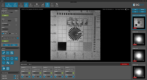
Sigray3D | Intuitive Acquisition
- Easy, intuitive software gets your team up and running in no time
- Click to align the sample and start measuring in seconds
- AI powered AutoPilot suggests the optimal settings for each sample
- Queue up to 20 samples with the automated Sample Handling Robot (SHR)
Image 1: GigaRecon provides high quality data with far fewer views required
Image 2: Sigray3D features an intuitive GUI for acquiring and measuring data
SPECIFICATIONS
| Parameter | ||
|---|---|---|
| Overall | Spatial Resolution | Minimum 35nm spatial resolution at 2,7 keV |
| Source | Type(s) | Patented multi-target reflection source |
| Voltage | 20-50 kVp | |
| Target(s) | Up to 3 targets. Si, Cr, Fe, Cu |
|
| Modes | Resolution and FOV Settings | 3 high resolution modes and 1 LFOV mode |
| LFOV Mode | 120nm resolution | 65nm voxel 65 μm FOV | 60 μm diameter optimal |
|
| Optics | Condensers | 3 sets of Sigray Double Paraboloidal x-ray optics matched to zone plates |
| Focusing Objective | Fresnel diffraction zone plates | |
| Phase Ring | Zernike phase shift ring | |
| Software | Command and Control | Sigray 3D with Intuitive interface |
| Reconstruction | GigaRecon – fastest commercial CBCT reconstruction software | |
| Linux Workstation | Interface is on a Windows workstation, while a separate robust Linux workstation controls the system. Advantageous for reliable 24-7 operation. | |
| EPICS | Open-source software controls for maximum flexibility | |
| Dimensions | Footprint | 2.3m L x 1.3m W x 1.5m H |


