


Prof. Kirkland is the Science Director at the new Rosalind Franklin Institute, UK
Courtesy of Olaf Weller, DENSsolutions
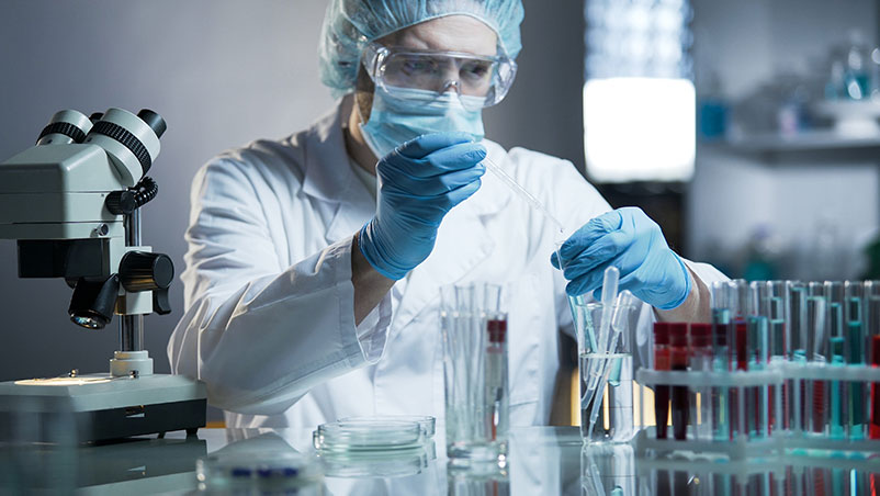
DENSsolutions interviewed Prof. Angus Kirkland, Professor at the Department of Materials, University of Oxford and the science director at the Electron Physical Science Imaging Centre (EPSIC), Diamond Light Source UK. They talked about the new Rosalind Franklin Institute where Prof.Kirkland performs disruptive research projects in life sciences involving physical science methods, techniques, and instruments including In Situ TEM and correlative imaging.
Prof. Kirkland discussed his research with colleagues at UCL involving Brownian tomography and with colleagues in Oxford looking at defect dynamics in low dimensional materials like graphene. With his diverse experience in TEM, Kirkland discusses cutting-edge ideas on future advancements in liquid-phase electron microscopy (LPEM) and examines the way that TEM research and big data mining are becoming intertwined.
“What we would like to ultimately aim for is to be able to image important biological structures in their native environment; so in aqueous solution at about 37.5°C, while interacting with other biological structures or pharmaceutical compounds with timing resolutions of about a microsecond. ”
How did you get involved with In Situ TEM research?
“Well, I originally got involved in TEM research because my PhD was looking at the structures of small metal particles and any broad beam technique at that time just simply gave you structural averages. So, the only obvious methodology was to use a microscopy-based technique and TEM was the obvious choice. So, I got heavily involved in high-resolution TEM as part of my PhD.
I then spent a lot of time developing methods for TEM, including super-resolution methods, and we got back into In Situ TEM when I moved to Oxford because we were then interested in mapping the phase diagrams (the structural changes as a function of temperature and size) for small metal particles. So, we contacted DENSsolutions in the very early days when there were only a few people working here, and we purchased one of the very early In Situ heating holders and published some very nice papers on the phase diagram for nanogold.”
How did the DENSsolutions system aid your In Situ research?
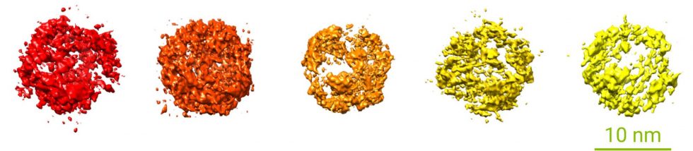
“The DENSsolutions system gave us much better drift stability and temperature control than any other product in the market. We would take a small gold particle and map out how its structure changed as a function of temperature very accurately, or we could take particles of different sizes. So Dr. Neil P. Young, University of Oxford, UK, and I actually mapped out the phase diagrams experimentally and compared them to theoretical calculations, done by Dr. Amanda Barnard, CSIRO, Australia, of what the predicted phase diagram would be. And they mapped almost perfectly.”
Did this research make changes in industry?
“This was a critically important problem for the catalysis industry because gold is used for carbon monoxide oxidation as a catalyst. The catalysis industry would like to know: if we have a gold particle of a certain size; what’s its structure, then what’s its surface structure, and what’s its catalytic activity? And the traditional way is to do lots of experiments; you would measure thousands of particles experimentally which takes time.
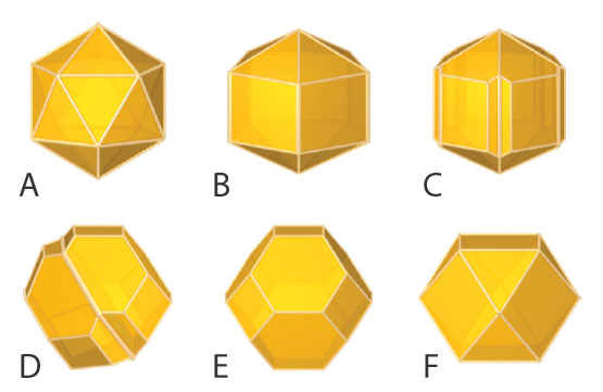
Reprinted with permission from Barnard et al, Jun 2, 2009, ACS Nano, doi.org/10.1021/nn900220k. Copyright 2009 American Chemical Society.
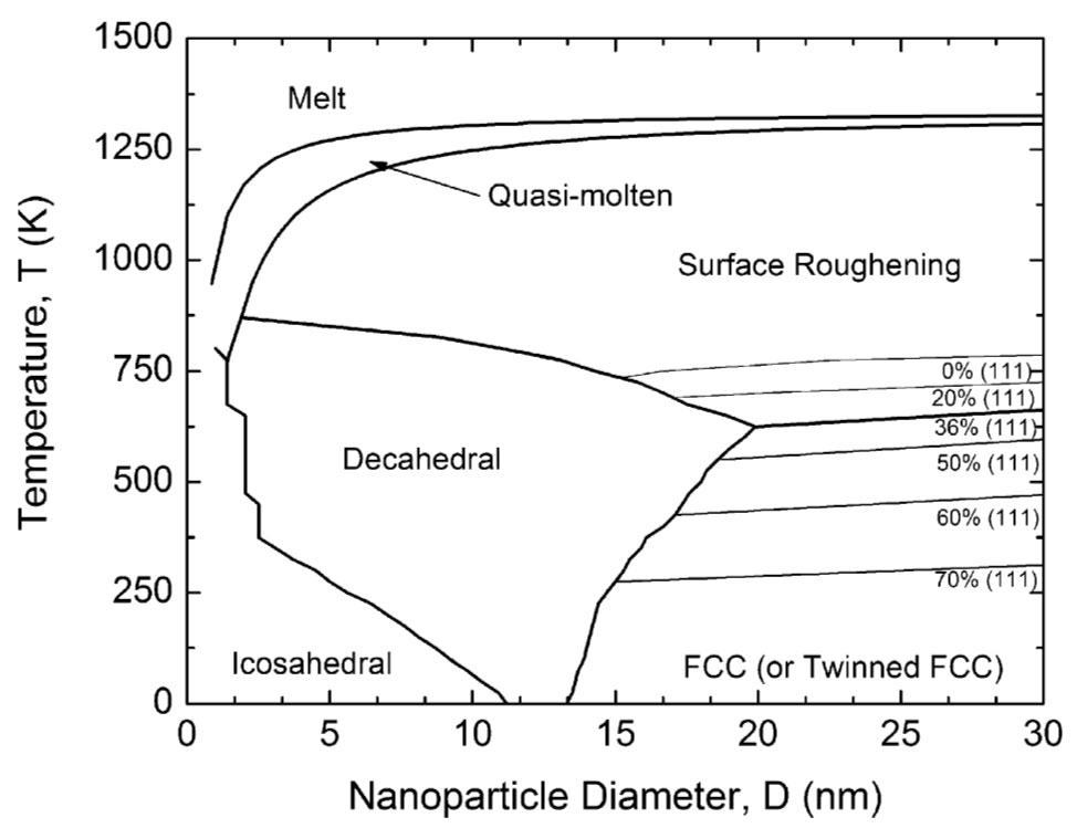
Reprinted with permission from Barnard et al, Jun 2, 2009, ACS Nano, doi.org/10.1021/nn900220k. Copyright 2009 American Chemical Society.
What they do now is they take our phase diagram (Fig. 2.) and they say: so if it’s 30 nm at room temperature it will have this shape. So, they can use it as a predictive tool to understand how best to optimise their catalysts. I’m very proud of that paper because it means that industry doesn’t have to do thousands of experiments. They can take one diagram and use its predictive power. link1 link2.”
Which other In Situ TEM solutions from DENSsolutions have you been using?
“I’ve got a student at the moment, he’s actually the Rhodes Mandela scholar in Oxford, and he’s been looking at Fischer Tropsch catalysts which are complicated cobalt or iron metal-and-metal oxide systems. We’ve been using the DENSsolutions Climate system extensively to do In Situ Fischer Tropsch catalysis and mapped that data back onto the very large studies that have been done ex situ to verify that the ex situ studies and the ex situ microscopy are correlated.
Most recently, I have become very interested in liquid cells for developing imaging methods to look at biological macromolecules and biological structures In Situ in liquid environments rather than in frozen and vitrified ice. That’s a driver in the new Rosalind Franklin Institute for which I am one of the science directors.”

Can you tell us about the Rosalind Franklin Institute?
“The original concept for the Rosalind Franklin Institute was initiated by Sir John Bell at Oxford University. A team of people were assembled across different disciplines, including myself, to set up the initial science themes. At that point, I was invited to lead the theme ‘Correlated Imaging’. We had to write the science case, that was peer-reviewed, then we had to write the business case that was also reviewed by government, and then it was funded, so I was involved from day one.
The Rosalind Franklin Institute’s main mission statement is to “translate physical science methods, techniques and instruments across the boundary into life sciences and medicine.” To do this I had to learn very quickly a bit of biology “101” and start understanding life sciences. So, that’s why I got interested in life sciences. I’m a chemist/physicist by background and now I have the unique opportunity to take all that I have learned and apply it to the field of life science.”
Is this also how you got interested in liquid-phase electron microscopy (LPEM)?
“Yes, absolutely. One of the things that we will be doing in the Rosalind Franklin Institute with the liquid cell developments is trying to apply some of the methods that we conventionally use in vacuum into the liquid state. So, we’ve developed a lot over the years; various phase retrieval methods, including ptychographic experiments and we’d like to try all of those, not in vacuum, but in the liquid state.
Brownian tomography (developed at UCL by Prof. Giuseppe Battaglia and his group), is a powerful technique that can be used to reconstruct a 3D model without tilting the holder. This is one of the techniques we would like to apply but there are also other experiments which we know how to do in a vacuum and we’d like to see how they translate into the liquid state. This will give us extra additional information that we otherwise wouldn’t have had.”
Which of the projects inside the Rosalind Franklin Institute are you most excited about?
“The Rosalind Franklin’s mission is to do disruptive science. What we would like to ultimately aim at is to image important biological structures in their native environment, so in aqueous solution at about 37.5°C, while interacting with other biological structures or pharmaceutical compounds with timing resolutions of about a microsecond. So, making million-frames per second movies of images of biology in action: That’s the moonshot.

For this, we need further developments in liquid cell technology. We need MEMS devices holding liquids at body temperature very accurately where you can actually flow in, for example, a saline solution or a sugar solution, or even a dilute solution of a pharmaceutical compound. In order to do anything meaningful in this field we have to have incredibly low drift rates, we have to have very accurate flow control and we have to be able to deal with liquids that are slightly viscous so that’s an engineering challenge in itself, and finally, of course, we have to have accurate heating control.
Basically, we’d like a liquid cell that mimics the human body.”
What is needed to reach this goal?
“There’s a lot of engineering to be done in terms of not only the liquid cell holder development but also in terms of the instrumentation to give us the very fast beam blanking and the new electron optical columns that we’re going to need. We have JEOL as a partner to do that with. Next, we also need to think carefully about how we’re going to manage and mine the data because we’re going to generate huge quantities of data very quickly, finally we have to upgrade and develop a new generation of electron detectors.
So: engineering, detector physics liquid cell development and electron optics all need to be advanced.”
How will other collaborators, like DENSsolutions, contribute to the research?
“In terms of liquid cell development, we need a reliable commercial manufacturer like DENSsolutions because we don’t do the precision engineering and the MEMS design. The column optics and the fast shuttering will be done by JEOL and partners who specialise in that area.
The team we will need to put together internally is going to be a combination of engineers and scientists and computational and data scientists for managing the data. We will almost certainly need to hire some theorists to deal with the necessary algorithms that we have to develop, for example, for aligning and stacking the data.
We will of course need to have biologists in that team to identify some really relevant early biological problems that we can tackle. So the whole point of the Rosalind Franklin Institute is to assemble these very multidisciplinary teams all attacking one big long-range problem.”
Do you see a merging of materials science and life science happening?
“Yes, this is one of the things we have already found at the national centres. We have two national centres, one in physical sciences and one in life sciences (ePSIC and eBIC), and it’s the former that I direct. Because we have life scientists and physical scientists already sitting in the same building on adjacent desks, we’ve already found that there’s been a huge collaborative interaction between them.
So, there’s been a really nice convergence of our interests and that’s actually led to a couple of very nice experimental programs which we wouldn’t have thought about without their input. We’ve got a couple of publications going through at the moment with some new data.”
Can you tell us something about your recent work?
“I guess that most of the work that I’ve been doing recently has been in the materials science sector. I have had a very fruitful and successful collaboration with Prof. Jamie Warner in Oxford looking at defect structures in low dimensional materials link. We started with graphene, we looked at silicon nitride, we looked at various disulphides and that’s produced a string of very high impact papers over the last seven years or so.
The other paper we’re proud of, which unfortunately doesn’t involve In Situ at the moment, was that we solved a very old problem, in collaboration with Nelson Mandela University in South Africa. This was a fifteen-year-old problem that hadn’t been resolved: the structure of nitrogen-containing-platelets in natural type 1a diamonds. Natural type 1a diamonds have nitrogen-containing inclusions and the structure of those platelets has been a controversy that has been unresolved for the last fifteen years or so. We finally, unambiguously, determined which of the four models that have been proposed was correct link. So that was a very satisfying piece of work.”
Which of your work was enabled by DENSsolutions In Situ systems?
“Most of the In Situ work that we’ve done, in which we used the DENSsolutions Wildfire system extensively, has been looking at variable temperature defect formation and migration in these low dimensional materials.
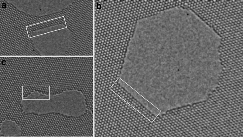
Reprinted with permission from Kuang He et al, April 16, 2015, ACS Nano, doi.org/10.1021/acsnano.5b01130. Copyright 2015 American Chemical Society.
One of the earliest things we did with the early DENSsolutions heating holders was to suspend sheets of graphene across FIB holes in the heating element and look at how defect dynamics changed as a function of temperature. So the In Situ part, i.e. the precise temperature control, was very critical. We are now extending that research with very fast detectors to look at much bigger data sets to try and extract the chemical kinetics of these defect formations and transformations.”
What do you expect from DENSsolutions in the future?
“I know that you are ahead of the game when it comes to the next generation of liquid cells and you already have a huge amount of technology that we’re going to need.
“It’s a question of maybe expanding that a little bit in certain key areas. I think the other thing we’d like, and this is something applicable to all the manufacturers, is to be able to move the specialist end components of the holder, the sample carrier, from an electron microscope for example into an X-ray beamline or an optical microscope.
We would ideally like a common platform standard so that we can move the same system between instruments because I think there is a huge amount of interesting correlation studies that can be done if you can image the same sample using lots of different imaging instruments. We would like to modularise the holder still further so that we’re not reliant on a specific type of TEM rod. If you could provide the In Situ platform which we can use in any correlated imaging workflow, that would be incredibly powerful.”
How is your research influenced by societal problems?
“Within the Rosalind Franklin Institute, it’s a core part of our mission because we’re translating physical science into life sciences and medicine, so a lot of the problems that we will attack are going to be pulled or pushed by clinicians and people working in medical research who need specific kinds of information.
For example, there are people with cancer who are treated with cisplatin drugs which are platinum-containing organometallic compounds. The problem is that sometimes people get a very acute pain when they have this type of chemotherapy and the questions are “why do they get the pain? Where does the platinum go?” This is a clinical problem that’s currently being explored using electron microscopy, which is a physical science method.”
So Prof. Peter Nellist, University of Oxford, UK, and his student Alex Schreader are looking at the cellular distribution of the platinum after the treatment in collaboration with King’s College London. We want to see where the platinum goes in relation to the cellular context, and does it differ for different drugs, for different types of patients, for different stages of treatment? There are lots of good medical problems that can be solved with electron microscopy but, of course, if you want to do anything medical which has a societal benefit you really have to do it In Situ in as close to a native state as possible.
The problem is that the native state isn’t ultra-high vacuum in an electron beam, it’s in a liquid environment or possibly a frozen environment. So that’s where the In Situ part becomes really relevant. Otherwise, it’s very hard to verify if what you see in ultra-high vacuum conditions is relevant to physiological environments.”
Which recent publication wowed you in the field In Situ TEM, outside of your own research?
“If I’m thinking about In Situ work, the publications that I’ve found most impressive are the In Situ experiments where people actually put real devices into the microscope and bias them and see how, for example, doping contributions change, how electric fields change. Here, you actually get a very accurate structural and chemical picture of a real device in operando.
You can observe, for instance, a genuine PN junction, bias it, and see how the field changes and where the dopants move to. It was nice to see these images, you look at the field lines and images and they look exactly like the textbook diagrams. There are a few groups doing this kind of work, for example the group of Prof. Raphal Dunin-Borkowski, ER-C Jülich, Germany, and Dr. Martin Hytch, CEMES-CNRS, France.
Other In Situ work that comes to mind is that of Prof. Frances M. Ross, DMSE, MIT US, who has done a lot of In Situ work looking at vapour-liquid deposition of various nanowires. She’s got fantastic movies of nanowires with gold caps at the top, showing nanowires growing out of a substrate and making forests link.
She’s also developed all the maths and calculations to work out the growth kinetics as a function of precursor concentrations.”
What are some of the advantages of working In Situ?
“An advantage of the In Situ work is really all about being able to model and visualise the formation of structures. So, you can put the component parts together in their natural environment, whatever that might be; a chemical environment, a gas environment, a liquid environment, and actually see how they assemble themselves into the final composite device. So, you’re actually seeing how things form rather than just looking at the post mortem structures in vacuum after they form.
An important part of the work is building models that predict how these would form so you can change components of that model building process and show how different structures would arise. In this way you can develop some sort of relationship between the component parts and the final structure and the final property which tailors your devices. So that’s where we and others are heading for.”
What will be the big challenges for TEM research overall in the coming years?
“I think one challenge for electron microscopy in the next few years is going to be the big data challenge. We’ve scaled up over the last few years from megabytes to gigabytes and now to terabytes, we’re now hitting the many-terabytes or even hundreds-of-terabytes limit, and in some areas towards petabytes. Then the whole data problem changes.

This is driven, in part by an increasing time resolution. With current detectors, for our graphene-defect work, we routinely feed into a neural network, developed by Dr. Chen Huang, one of my postdocs over one-and-a-half to two million images taken in one session. So, whereas one-and-a-half to two million images might be the total output from a research group in ten years, now it’s half a day. So were generating vast quantities of data.
The challenge is not just about storing it and archiving it, it’s actually about mining it. How do you extract the information you want from these very large datasets? It is clear to us that you can’t do that manually, so you have to turn to machine learning and artificial intelligence. So Chen has spent a lot of time recently developing AI and machine learning applied to specific EM problems with these large datasets.”


